. 2009 Nov 23;206(12):2735-45.
doi: 10.1084/jem.20090222. Epub 2009 Nov 9.

Key role of T cell defects in age-related vulnerability to West Nile virus

Affiliations

Key role of T cell defects in age-related vulnerability to West Nile virus

James D Brien et al. J Exp Med. .

Abstract

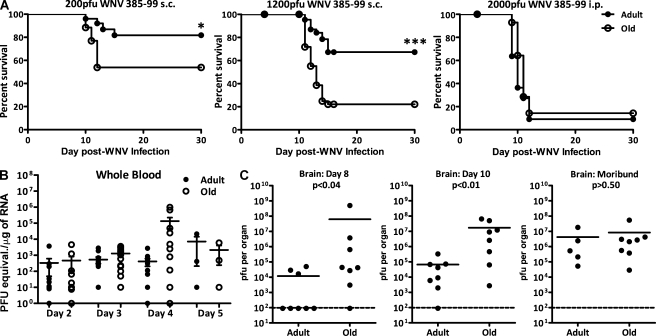
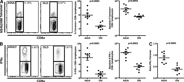
West Nile virus (WNV) infection causes a life-threatening meningoencephalitis that becomes increasingly more prevalent over the age of 50 and is 40-50x more prevalent in people over the age of 70, compared with adults under the age of 40. In a mouse model of age-related vulnerability to WNV, we demonstrate that death correlates with increased viral titers in the brain and that this loss of virus control with age was the result of defects in the CD4 and CD8 T cell response against WNV. Specific age-related defects in T cell responses against dominant WNV epitopes were detected at the level of cytokine and lytic granule production, each of which are essential for resistance against WNV, and in the ability to generate multifunctional anti-WNV effector T cells, which are believed to be critical for robust antiviral immunity. In contrast, at the peak of the response, old and adult T cells exhibited superimposable peptide sensitivity. Most importantly, although the adult CD4 or CD8 T cells readily protected immunodeficient mice upon adoptive transfer, old T cells of either subset were unable to provide WNV-specific protection. Consistent with a profound qualitative and quantitative defect in T cell immunity, old brains contained at least 12x fewer total effector CD8 T cells compared with adult mice at the peak of brain infection. These findings identify potential targets for immunomodulation and treatment to combat lethal WNV infection in the elderly.


Figures

Figure 1.
Figure 1.
Age-related susceptibility to WNV disease in old mice. (A) Survival of adult (4–6 mo old) and old (18–22 mo old) mice after challenge with the indicated doses of WNV 385–99 s.c. or i.p. Old mice are five and six times more susceptible to WNV upon i.p. (MST, 13 d) and s.c. (MST, 14 d) infection, respectively, compared with adult controls. Statistical significance was evaluated by the Log-rank test (***, P < 0.0005; *, P < 0.05). Please note that difference between the old and adult mice disappears at the high viral challenge dose, where essentially all animals succumb to infection, and also at the low virus dose, where old animals appear to control the low viral loads well (left; see also Table S1 for additional experiments with other viral isolates and routes). Results depict mortality (n = 12 mice/group) and are representative of at least four experiments with similar results. (B and C) Viral titers within indicated tissues and organs of adult and old mice after s.c. infection (1,200 PFU/mouse) were determined by quantitative PCR (B, whole blood expressed in approximate PFU equivalents, based on quantitative PCR of a known WNV RNA standard) or by plaque assay (C, brain). The brains of old mice contained significantly more virus on both day 7 and day 10 after infection. In contrast, there was no difference between brain viral levels of old and adult moribund mice on day 12–16. For data on day 3 blood and spleen by plaque assay, please see Fig. S2. Horizontal bars indicate mean values of the unpaired Student's t test. Error bars represent SEM.
Figure 2.
Figure 2.
Relative roles of innate and adaptive immunity and the importance of the age of T cells in resistance to WNV infection. (A) IFNAR−/−, RAG1−/−, and WT mice were infected with the indicated WNV doses s.c., and MST and percentage of survival were scored. WNV caused rapid mortality (MST, 4.5 d at 200 PFU) with a very low rate of survival in IFNAR−/− mice compared with WT controls (P < 0.0001 at 200 PFU). WNV infection also caused significant mortality in RAG-1−/− mice compared with WT controls (P < 0.0001) but MST was similar to that of WT mice (MST, 13 d at 200 PFU). Data were compiled from two independent experiments. (B) Adoptive transfer of lymphocytes from old and adult mice reveals age-related defects in adaptive immunity. Splenocytes (107/animal) from adult mice protect RAG-1−/− significantly better than splenocytes from old mice (top; MST, 14.5 at 100 PFU; P = 0.0025). Transfer of purified T cells (CD4 and CD8; 5 × 10⁶/mouse) reveals defects in old T cells, which were inferior at protecting RAG1−/− mice compared with the purified T cells from adult mice (bottom; 100 PFU; P < 0.03). Compared with RAG1−/− mice with no transfer, old splenocytes conferred no significant protection, whereas old T cells showed some protection (P = 0.04). **, P = 0.01–0.001; ***, P < 0.001. Results are representative of two comparable experiments.
Figure 3.
Figure 3.
Functional quantitative defects in T cell activation in response to WNV infection. Infection was as in Fig. 1 A. (A) CD8+ T cells derived from spleens of old and adult mice were harvested on day 8 and analyzed for the proportion (left and middle left, representative examples; middle right, aggregate analysis of relative numbers) and absolute numbers (right, absolute numbers) of cells expressing the CD8+ NS4b-2488:Kb+ phenotype. Dots denote individual mice (n = 8) and are representative of four experiments. (B) Same as A, except that measurements show relative representation and absolute numbers of CD8+ IFN-γ+ cells in response to the immunodominant NS4b-2488 CD8 epitope by ICCS (Brien et al., 2007) at the peak of the immune response (day 8). Old mice mobilized significantly fewer numbers of IFN-γ–producing WNV-specific CD8+ T cells than their adult counterparts. Results were compiled from three independent experiments. (C) Ratio between IFN-γ+ and Tet+ CD8+ cells shows that many fewer Ag-specific T cells are functionally able to manufacture this cytokine in the old mice. Groups, representation, and graphics are as in A, and results are representative of two experiments. Horizontal bars indicate mean values of the unpaired Student's t test. Error bars represent SEM.
Figure 4.
Figure 4.
Quality of WNV-specific responses in old mice is impaired at several levels. For all panels, animals were infected with 1,000 PFU WNV 385–99 and were analyzed on day 8, unless otherwise indicated. (A) At the peak of infection, splenic CD8 cells were stimulated with class I–restricted peptides and analyzed for the expression of IFN-γ, TNF, and GzB, and results were plotted to denote percentages of cells exhibiting all three molecules (3 Fxn), two molecules (2 Fxn), or a single molecule (1 Fxn). Results depict eight animals per group and are representative of two such experiments. (B) Relative intensity of expression of IFN-γ, TNF, and GzB among CD8+ cells in response to 6 h of NS4b-2488 peptide stimulation as described in Materials and methods. After gating on CD8+ T cells, normalized mean fluorescent intensity (MFI) was obtained by subtracting the mean fluorescent intensity of the negative cell population from that of the positive cells. Data depict five adult and six old individual mice, representative of four experiments, and are shown as in Fig. 1 A. (C) Expression of CD43 on WNV-specific CD8 cells from adult and old mice was analyzed on day 8 after infection by selective gating and is expressed as percentage of Tet+CD8+ cells that are CD43+, shown for eight animals per group and representative of three such experiments. Horizontal bars in B and C indicate mean values of the unpaired Student's t test. (D) Ex vivo cytotoxic activity of adult and old CD8+ T cells demonstrates major age-related functional defects. Adult and old mice were infected s.c. with WNV at a dose (1,000 PFU) that caused decreased survival of old mice compared with adult animals. CD8+ T cells from adult mice exhibited stronger cytotoxic activity than CD8+ T cells from old mice when assayed directly ex vivo 7 d after infection in a 6-h standard 51Cr-release assay. Peptide-coated NS4b2488 (10−6M) EL-4 cells were used as targets. Values for five mice per group with standard deviations, representative of two experiments, are shown. P-values are depicted above the graphs.
Figure 5.
Figure 5.
Inability of aged CD4 and CD8 T cells to protect RAG-1−/− mice against lethal WNV infection. (A) Purified CD8 and CD4 T cells from C57BL/6 (closed squares), IFNγ−/− (open squares), or perforin−/− (closed triangles) mice were transferred into RAG-1−/− mice, which were then infected with 200 PFU WNV and scored for survival. WT T cells exhibited significantly enhanced protection when compared with RAG-1−/− mice with no transfer (closed circles, P < 0.0006) or RAG-1−/− mice receiving perforin−/− (P < 0.003) or IFN-γ−/− T cells (P < 0.01). (B) Young RAG-1−/− mice received no cells (closed diamonds) or received highly purified adult (squares) or old (inverted triangles) CD4 (closed symbols) or CD8 (open symbols) T cells (5 × 10⁶ cells/mouse). Engraftment was verified after 24 h, with animals infected with 200 PFU WNV, and survival was scored thereafter. Adoptive transfer of old CD4 or CD8 T cells failed to confer any protection to RAG-1–deficient hosts, whereas transfer of adult CD4 (P < 0.0001) or CD8 (P < 0.01) T cells afforded a high degree of protection. Both parts of the figure were reproduced in three separate experiments.
Figure 6.
Figure 6.
Analysis of brain infiltrates from WNV-infected adult and old mice. The brains of old and adult C57BL/6 mice were harvested 12 d after infection with 1,000 PFU WNV 385–99 s.c., sectioned, and costained for CD3 (green) and WNV (red). (A) Representative adult (top) and old (bottom) brains are shown at medium (left) and high (right) power. CD3 T cells are stained green and WNV-infected neurons are red. Bar: (left) 50 µm; (right) 20 µm. (B) Brains of 10 old and 10 adult mice harvested 10 d after infection with WNV. To generate five independent samples per group, with sufficient cell numbers for analysis, pools of two brains each were made and cells isolated using percoll gradient. The representation of cells using mononuclear/lymphocyte gate in representative adult (left) and old (middle) mice are shown. The aggregate analysis of cell percentages in this gate are shown on the right, illustrating that there are significantly fewer cells in the lymphocyte gate in old mice. Comparison is based on at least 5 × 10⁵ collected events/sample. (C) Representation (left) and aggregate analysis (right) of the percentage of CD4 and CD8 T cells within the brain of old versus adult mice as determined by flow cytometry. Significant differences were seen in representation of these cell subsets as well. (D, left) Representation of CD8 NS4b-2488 tetramer+ T cells among all CD8 T cells in the brains of old mice is further reduced compared with adults (P = 0.00372). (D, right) Illustration of cumulative effects of reduced representation of total lymphocytes, CD8 cells, and CD8 NS4b-2488 Tet+/GrB+ T cells in the brains of old mice compared with adults (taken as 100%) reveal an ∼12× age-related difference. Experiment is representative of three independent experiments. Horizontal bars indicate mean values of the unpaired Student's t test. Error bars represent SEM.

References

    1. Anderson J.F., Andreadis T.G., Vossbrinck C.R., Tirrell S., Wakem E.M., French R.A., Garmendia A.E., Van Kruiningen H.J. 1999. Isolation of West Nile virus from mosquitoes, crows, and a Cooper's hawk in Connecticut. Science. 286:2331–2333 10.1126/science.286.5448.2331 - DOI - PubMed
    1. Bender B.S., Taylor S.F., Zander D.S., Cottey R. 1995. Pulmonary immune response of young and aged mice after influenza challenge. J. Lab. Clin. Med. 126:169–177 - PubMed
    1. Brien J.D., Uhrlaub J.L., Nikolich-Žugich J. 2007. Protective capacity and epitope specificity of CD8(+) T cells responding to lethal West Nile virus infection. Eur. J. Immunol. 37:1855–1863 10.1002/eji.200737196 - DOI - PubMed
    1. Brien J.D., Uhrlaub J.L., Nikolich-Žugich J. 2008. West Nile virus-specific CD4 T cells exhibit direct antiviral cytokine secretion and cytotoxicity and are sufficient for antiviral protection. J. Immunol. 181:8568–8575 - PMC - PubMed
    1. Cambier J. 2005. Immunosenescence: a problem of lymphopoiesis, homeostasis, microenvironment, and signaling. Immunol. Rev. 205:5–6 10.1111/j.0105-2896.2005.00276.x - DOI - PubMed

Publication types

MeSH terms