. 2009 Nov 24;120(21):2077-87.
doi: 10.1161/CIRCULATIONAHA.109.884403. Epub 2009 Nov 9.

Enhancement of myocardial regeneration through genetic engineering of cardiac progenitor cells expressing Pim-1 kinase

Affiliations

Enhancement of myocardial regeneration through genetic engineering of cardiac progenitor cells expressing Pim-1 kinase

Kimberlee M Fischer et al. Circulation. .

Abstract

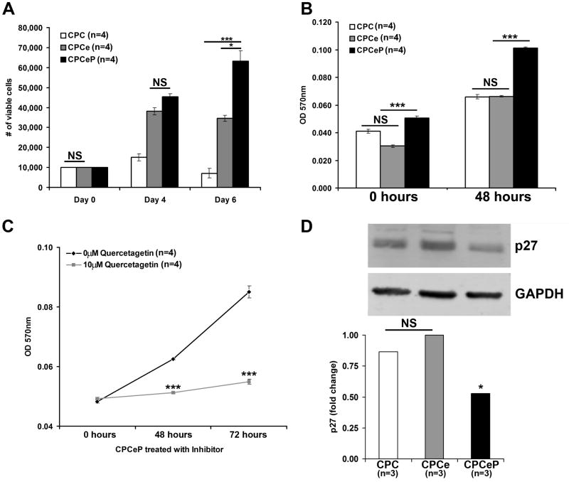
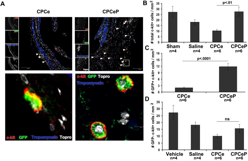
Background: Despite numerous studies demonstrating the efficacy of cellular adoptive transfer for therapeutic myocardial regeneration, problems remain for donated cells with regard to survival, persistence, engraftment, and long-term benefits. This study redresses these concerns by enhancing the regenerative potential of adoptively transferred cardiac progenitor cells (CPCs) via genetic engineering to overexpress Pim-1, a cardioprotective kinase that enhances cell survival and proliferation.

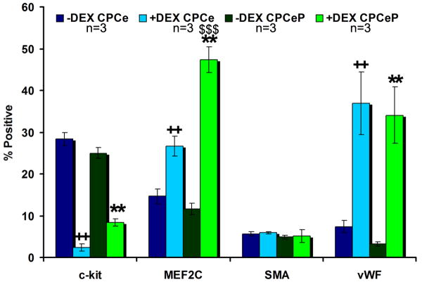
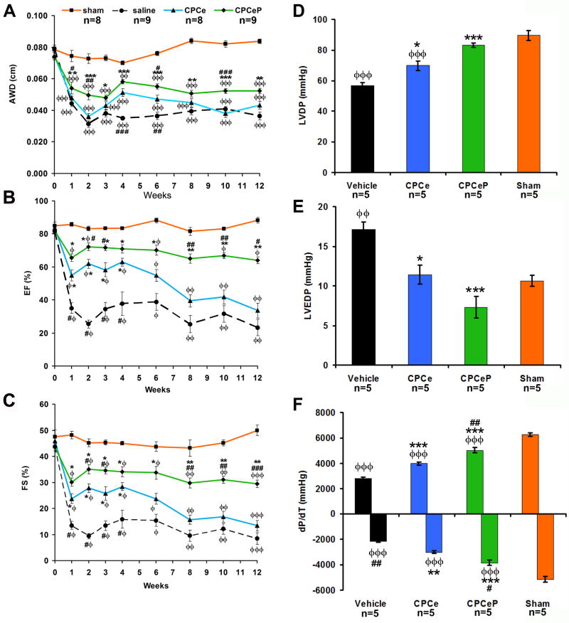
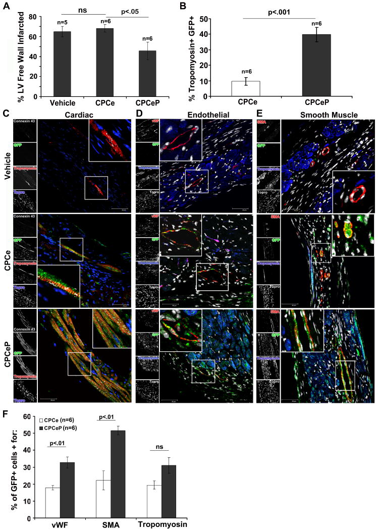
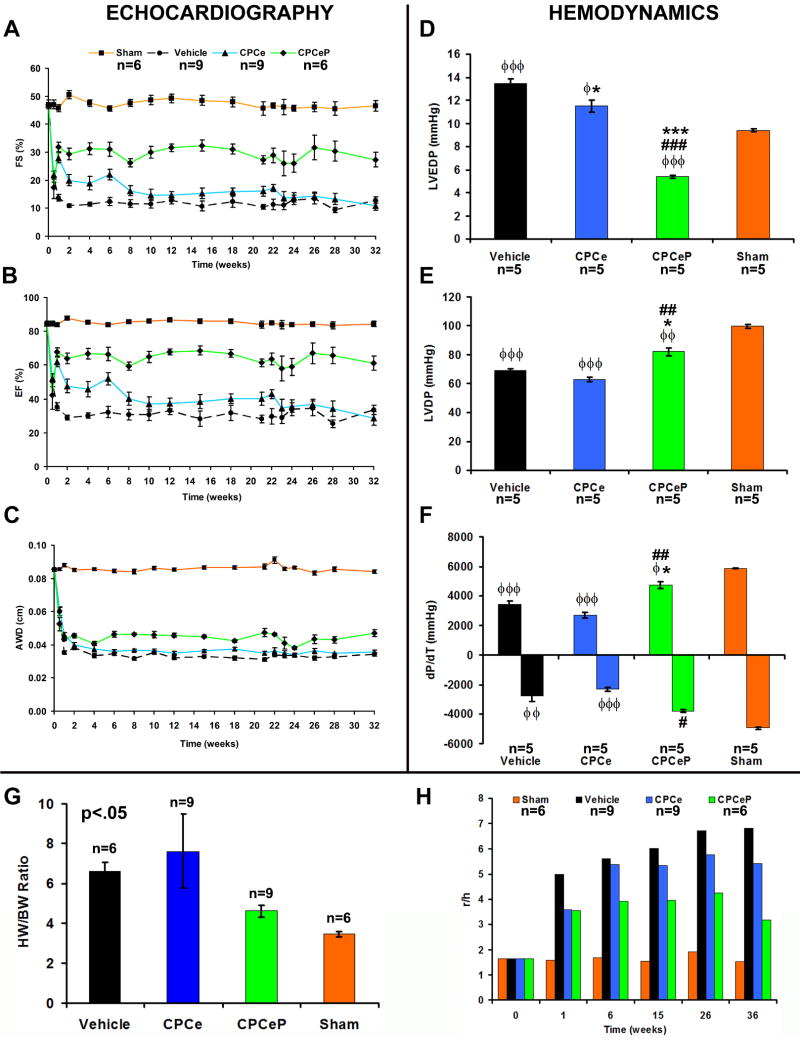
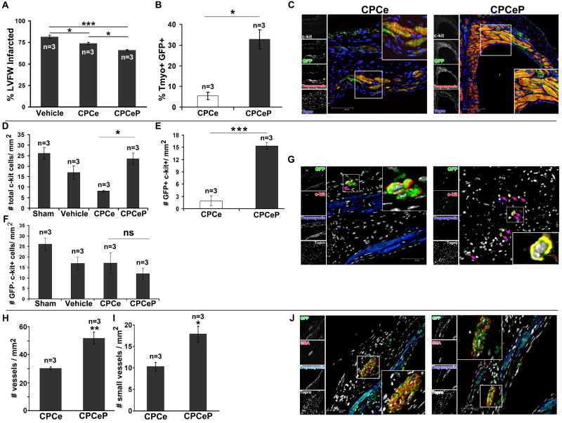
Methods and results: Intramyocardial injections of CPCs overexpressing Pim-1 were given to infarcted female mice. Animals were monitored over 4, 12, and 32 weeks to assess cardiac function and engraftment of Pim-1 CPCs with echocardiography, in vivo hemodynamics, and confocal imagery. CPCs overexpressing Pim-1 showed increased proliferation and expression of markers consistent with cardiogenic lineage commitment after dexamethasone exposure in vitro. Animals that received CPCs overexpressing Pim-1 also produced greater levels of cellular engraftment, persistence, and functional improvement relative to control CPCs up to 32 weeks after delivery. Salutary effects include reduction of infarct size, greater number of c-kit(+) cells, and increased vasculature in the damaged region.

Conclusions: Myocardial repair is significantly enhanced by genetic engineering of CPCs with Pim-1 kinase. Ex vivo gene delivery to enhance cellular survival, proliferation, and regeneration may overcome current limitations of stem cell-based therapeutic approaches.


Conflict of interest statement

Disclosures: None

Figures

Figure 1
Figure 1. Pim-1 increases the proliferation rate of CPCs in vitro
(A) The number of viable cells as determined by trypan blue exclusion was determined for CPCeP, CPCe, and CPCs over a six day time course (mean ± SEM, n=4/group). (B) Metabolic rate of CPCeP, CPCe, and CPCs was measured by MTT assay over a 48 hour time course (mean ± SEM, n=4/group). (C) Metabolic rate evaluated by MTT assay in CPCeP treated with and without 10μM Quercetagetin over a 72 hour time course (mean ± SEM, n=4/group). (D) Immunoblot analysis of p27 protein in CPCeP, CPCe, and CPCs (n=3/group). *p<.05, **p<.01, ***p<.001.
Figure 2
Figure 2. Flow cytometric analysis of CPCe and CPCeP
Analysis of cardiogenic lineages of CPCe and CPCeP before (dark blue and dark green) and after (light blue and light green) dexamethasone treatment (mean ± SEM, n=3/group). ++p<.01 compared to CPCe –Dex. **p<.01 compared to CPCeP -DEX, $$$p<.001 compared to CPCe +Dex.
Figure 3
Figure 3. Intramyocardial injection of CPCeP improves cardiac function 12-weeks post infarction
(A-C) Electrocardiographic assessment of AWD (A), EF (B), and FS (C), in sham (■, orange, n=8), vehicle (●, black, n=9), CPCe (▲, blue, n=8), and CPCeP (◆, green, n=9), 12-weeks post-infarction (mean ± SEM). (D-F) Cardiac function of sham (orange, n=5), vehicle (black, n=5), CPCe (blue, n=5), and CPCeP (green, n=5) were evaluated using in vivo hemodynamic measurements of LVDP (D), LVEDP (E), and dP/dT (F) 12-weeks post-intramyocardial injection (mean ± SEM). φp<.05, φφp<.01, φφφp<.001 compared to sham; #p<.05, ##p<.01, ###p<.001 compared to vehicle, * p<.05, **p<.01, ***p<.001 compared to CPCe. Echocardiography significant (p<.05) by two-way repeated measures ANOVA.
Figure 4
Figure 4. De novo myocyte formation and neovascularization results in reduction of infarct size in CPCeP treated animals
(A) Quantitation of infarction area in vehicle (n=5), CPCe (n=6), and CPCeP (n=6) treated hearts 12-weeks post injection (mean ± SEM). (B) Quantitation of Tropomyosin+ eGFP+ cells in CPCe (n=6) and CPCeP (n=6) animals (mean ± SEM). (C-E) Representative immunostaining for colocalization of eGFP (green) and cardiac myocytes (tropomyosin, red) and connexin-43 (white) (C), endothelial cells (vWF, red) (D), and smooth muscle cells (SMA, red) (E) in vehicle, CPCe, and CPCeP treated hearts 12-weeks post-intramyocardial injection. Enlarged areas are represented with indicated boxes. Scale bars represent 40μm. (F) Quantitation of eGFP+ population in CPCe (n=6) and CPCeP (n=6) for cardiac lineage markers.
Figure 5
Figure 5. CPCeP treated hearts have increased numbers of c-kit+ cells 12-weeks post intramyocardial injection
(A) Immunostaining for c-kit and eGFP (green) in heart sections from mice treated with CPCe (n=6) or CPCeP (n=6) 12-weeks post-intramyocardial injection. Enlarged areas are represented with indicated boxes. (B) Quantitation of the number of total, (C) eGFP+, and (D) eGFP- c-kit+ cells in hearts of mice injected with CPCe (n=6) or CPCeP (n=6) (mean ± SEM). Scale bars represent 120μm in top panels.
Figure 6
Figure 6. Long-term persistent cardiac functional recovery in animals treated with CPCeP
(A-C) Electrocardiographic assessment of FS (A), EF (B), and AWD (C), in sham (■, orange, n=6), vehicle (●, black, n=9), CPCe (▲, blue, n=9), and CPCeP (◆, green, n=6), 32-weeks post-infarction (mean ± SEM). Statistics for each time point are provided in Table S1. (D-F) Cardiac function of sham (n=5), vehicle (n=5), CPCe (n=5), and CPCeP (n=5) were evaluated using in vivo hemodynamic measurements of LVEDP (D), LVDP (E), and dP/dT (F), 32-weeks post-intramyocardial injection (mean ± SEM). (G) Heart weight: body weight ratios. Statistically significant p<.05 (ANOVA). (H) Wall stress assessment comparing ratio of left ventricular diameter (r) to wall thickness (h) from sham (n=6), vehicle (n=9), CPCe (n=9), and CPCeP (n=6) treated animals (mean ± SEM). φp<.05, φφp<.01, φφφp<.001 compared to sham; #p<.05, ##p<.01, ###p<.001 compared to vehicle, * p<.05, **p<.01, ***p<.001 compared to CPCe. Echocardiography significant (p<.05) by two-way repeated measures ANOVA.
Figure 7
Figure 7. Persistent engraftment and differentiation of Pim-1 expressing CPCs 32-weeks post intramyocardial injection
(A) Infarct size measurement, (B) quantitation of Tropomyosin+ eGFP+ cells, (C) and confocal micrographs of hearts injected with CPCe (n=3) or CPCeP (n=3) 32-weeks post-infarction. (mean ± SEM). Sections were stained for eGFP (green), Tropomyosin , c-kit (white), and nuclear stain Topro-3-iodide (blue). Quantitation of total (D), eGFP+ (E), and eGFP- (F) c-kit+ cells in heart sections from mice treated with CPCe (n=3) or CPCeP (n=3) 32-weeks post-infarction. (G) Immunolabeling with eGFP (green), c-kit , tropomyosin (blue), and nuclear stain Topro-3-iodide (white). (H) Quantitation of total number of vessels, (I) small vessels and (J) immunolabeling for vasculature in hearts from mice treated with CPCe (n=3) or CPCeP (n=3) 32-weeks post-infarction. Sections were stained with eGFP (green), smooth muscle actin , tropomyosin (blue), and nuclear stain Topro-3-iodide (white). Mean ± SEM, n=3, * p<.05, **p<.01, ***p<.001. Scale bars represent 50μm.

References

    1. Linke A, Muller P, Nurzynska D, Casarsa C, Torella D, Nascimbene A, Castaldo C, Cascapera S, Bohm M, Quaini F, Urbanek K, Leri A, Hintze TH, Kajstura J, Anversa P. Stem cells in the dog heart are self-renewing, clonogenic, and multipotent and regenerate infarcted myocardium, improving cardiac function. Proc Natl Acad Sci U S A. 2005;102:8966–8971. - PMC - PubMed
    1. Beltrami AP, Barlucchi L, Torella D, Baker M, Limana F, Chimenti S, Kasahara H, Rota M, Musso E, Urbanek K, Leri A, Kajstura J, Nadal-Ginard B, Anversa P. Adult cardiac stem cells are multipotent and support myocardial regeneration. Cell. 2003;114:763–776. - PubMed
    1. Leri A, Kajstura J, Anversa P, Frishman WH. Myocardial regeneration and stem cell repair. Curr Probl Cardiol. 2008;33:91–153. - PubMed
    1. Gonzalez A, Rota M, Nurzynska D, Misao Y, Tillmanns J, Ojaimi C, Padin-Iruegas ME, Muller P, Esposito G, Bearzi C, Vitale S, Dawn B, Sanganalmath SK, Baker M, Hintze TH, Bolli R, Urbanek K, Hosoda T, Anversa P, Kajstura J, Leri A. Activation of cardiac progenitor cells reverses the failing heart senescent phenotype and prolongs lifespan. Circ Res. 2008;102:597–606. - PubMed
    1. Fransioli J, Bailey B, Gude NA, Cottage CT, Muraski JA, Emmanuel G, Wu W, Alvarez R, Rubio M, Ottolenghi S, Schaefer E, Sussman MA. Evolution of the c-kit-positive cell response to pathological challenge in the myocardium. Stem Cells. 2008;26:1315–1324. - PMC - PubMed

Publication types

Substances