

The Standard Care vs Corticosteroid for Retinal Vein Occlusion (SCORE) study system for evaluation of optical coherence tomograms: SCORE study report 4
- PMID: 19901211
- PMCID: PMC2788490
- DOI: 10.1001/archophthalmol.2009.277
The Standard Care vs Corticosteroid for Retinal Vein Occlusion (SCORE) study system for evaluation of optical coherence tomograms: SCORE study report 4
Abstract
Objective: To describe grading procedures for optical coherence tomographic (OCT) images of participants in the Standard Care vs Corticosteroid for Retinal Vein Occlusion (SCORE) Study.
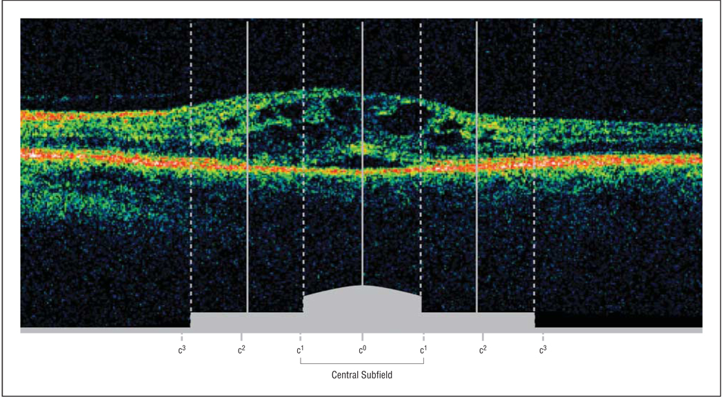
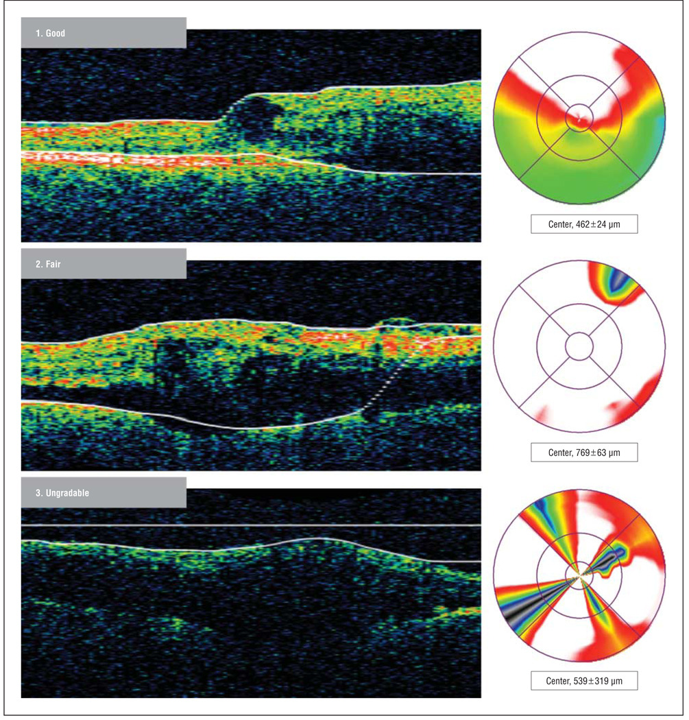
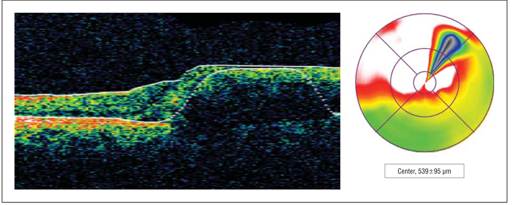
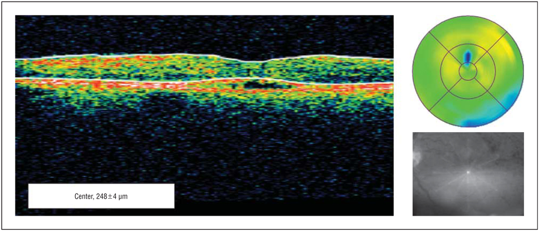
Methods: Optical coherence tomograms were taken at clinical sites with the Stratus OCT using fast macular and crosshair scan protocols. Paper prints of images were evaluated at a central reading center. Quality evaluation identified the accuracy of OCT-measured retinal thickness data and was categorized as good, fair, borderline, or ungradable. Manual measurement of center point thickness was performed on borderline images. Morphological evaluation identified cystoid spaces, subretinal fluid, and vitreoretinal interface abnormalities. Reproducibility of grading was assessed through formal quality control exercises.
Results: A randomly selected set of 106 images was identified for quality control. The first 2 annual regrades showed 91% and 89% intergrader agreement for OCT quality. Intraclass correlation for manually measured center point thickness was 0.99 per year. For morphological variables, intergrader agreement for cystoid spaces was 83% and 76%. Reproducibility for subretinal fluid and vitreoretinal interface abnormalities could not be interpreted owing to their limited presence in the sample.
Conclusion: Optical coherence tomogram evaluation procedures used in the SCORE Study are reproducible and can be used for multicenter longitudinal studies of retinal vein occlusion.
Figures

References
-
- Puliafito CA, Hee MR, Lin CP, et al. Imaging of macular diseases with optical coherence tomography. Ophthalmology. 1995;102(2):217–229. - PubMed
-
- Costa RA, Jorge R, Calucci D, Melo LA, Jr, Cardillo JA, Scott IU. Intravitreal bevacizumab (Avastin) for central and hemicentral retinal vein occlusions: IBeVO study. Retina. 2007;27(2):141–149. - PubMed
-
- Williamson TH, O’Donnell A. Intravitreal triamcinolone acetonide for cystoid macular edema in nonischemic central retinal vein occlusion. Am J Ophthalmol. 2005;139(5):860–866. - PubMed
