

Clinical syndromes associated with posterior atrophy: early age at onset AD spectrum
- PMID: 19901249
- PMCID: PMC2777069
- DOI: 10.1212/WNL.0b013e3181c0d427
Clinical syndromes associated with posterior atrophy: early age at onset AD spectrum
Abstract
Objective: Posterior cortical atrophy (PCA) and logopenic progressive aphasia (LPA) are clinical syndromes associated with posterior brain atrophy. We compared PCA and LPA to each other and to an age-matched group of patients with early age at onset of Alzheimer disease (EO-AD). We hypothesized that these 3 syndromes are part of a single clinical and biologic continuum.
Methods: Voxel-based morphometry (VBM) was used to assess atrophy in 14 PCA, 10 LPA, and 16 EO-AD patients compared to 65 healthy controls. Genetic analysis for APOE was conducted in 30 patients and 44 controls. Four patients came to autopsy. An additional 14 were studied with the beta-amyloid specific PET with tracer (11)C-labeled Pittsburgh Compound-B (PIB).
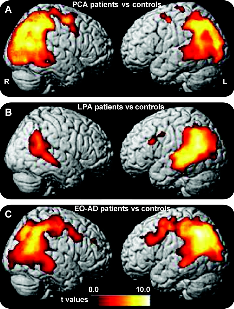
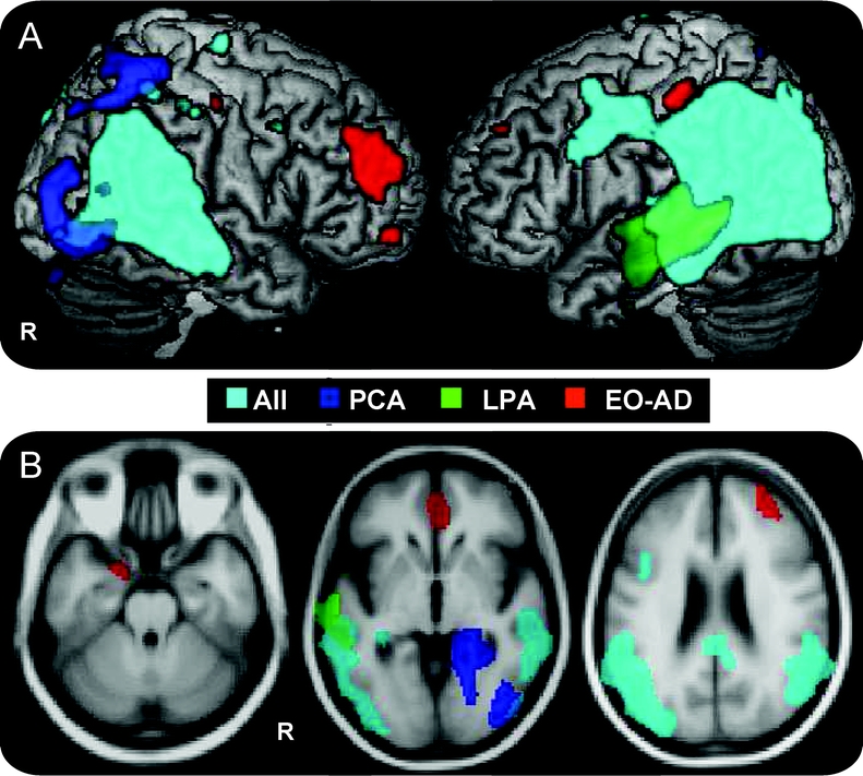
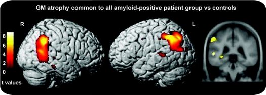
Results: VBM results demonstrated that, compared to controls, each patient group showed a large area of overlapping atrophy in bilateral parietal, occipital, precuneus, posterior cingulate, posterior temporal, and hippocampal regions. Surrounding this common area, group-specific atrophy was found in small, symptom-specific regions for each group: the right ventral-occipital and superior parietal regions in PCA, the left middle and superior temporal gyri in LPA, and the prefrontal cortex in EO-AD. APOE epsilon4 frequency was higher in all patient groups compared to controls. Four PCA, 5 LPA, and 8 EO-AD patients showed evidence of cortical amyloid at pathology (n = 3) or on PIB-PET (n = 14).
Conclusions: Logopenic progressive aphasia and posterior cortical atrophy showed largely overlapping anatomic and biologic features with early age at onset of Alzheimer disease, suggesting that these clinical syndromes represent the spectrum of clinical manifestation of the nontypical form of Alzheimer disease that presents at an early age.
Figures
Comment in
-
Clinical syndromes associated with posterior atrophy: early age at onset AD spectrum.Neurology. 2010 Aug 3;75(5):479; author reply 479-80. doi: 10.1212/WNL.0b013e3181e794d5. Neurology. 2010. PMID: 20679643 No abstract available.
References
-
- Tang-Wai DF, Graff-Radford NR, Boeve BF, et al. Clinical, genetic, and neuropathologic characteristics of posterior cortical atrophy. Neurology 2004;63:1168–1174. - PubMed
-
- von Gunten A, Bouras C, Kovari E, Giannakopoulos P, Hof PR. Neural substrates of cognitive and behavioral deficits in atypical Alzheimer’s disease. Brain Res Rev 2006;51:176–211. - PubMed
-
- Alladi S, Xuereb J, Bak T, et al. Focal cortical presentations of Alzheimer’s disease. Brain 2007;130:2636–2645. - PubMed
Publication types
MeSH terms
Grants and funding
LinkOut - more resources
Full Text Sources
Medical
Miscellaneous