Comparative Study
. 2010 Mar;58(3):237-46.
doi: 10.1369/jhc.2009.954321. Epub 2009 Nov 9.

The impact of tissue fixatives on morphology and antibody-based protein profiling in tissues and cells

Affiliations
Comparative Study

The impact of tissue fixatives on morphology and antibody-based protein profiling in tissues and cells

Linda Paavilainen et al. J Histochem Cytochem. 2010 Mar.

Abstract

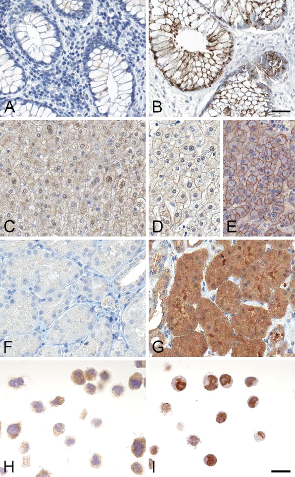
Pathology archives harbor large amounts of formalin-fixed, paraffin-embedded tissue samples, used mainly in clinical diagnostics but also for research purposes. Introduction of heat-induced antigen retrieval has enabled the use of tissue samples for extensive immunohistochemical analysis, despite the fact that antigen retrieval may not recover all epitopes, owing to alterations of the native protein structure induced by formalin. The aim of this study was to investigate how different fixatives influence protein recognition by immunodetection methods in tissues, cell preparations, and protein lysates, as compared with formalin. Seventy-two affinity-purified polyclonal antibodies were used to evaluate seven different fixatives. The aldehyde-based fixative Glyo-fixx proved to be excellent for preservation of proteins in tissue detected by immunohistochemistry (IHC), similar to formalin. A non-aldehyde-based fixative, NEO-FIX was superior for fixation of cultured cells, in regard to morphology, and thereby also advantageous for IHC. Large variability in the amount of protein extracted from the differently fixed tissues was observed, and the HOPE fixative provided the overall highest yield of protein. In conclusion, morphological resolution and immunoreactivity were superior in tissues fixed with aldehyde-based fixatives, whereas the use of non-aldehyde-based fixatives can be advantageous in obtaining high protein yield for Western blot analysis. This manuscript contains online supplemental material at http://www.jhc.org. Please visit this article online to view these materials.


Figures

Figure 1
Figure 1
Morphological detail was investigated using hematoxylin-eosin (H&E) staining. Superior morphology was observed with H&E in tissues fixed with aldehyde-based fixatives [neutral-buffered formalin (NBF) and Glyo-fixx], e.g., NBF in tonsil (A), whereas some shrinkage of cytoplasm is observed with the non-aldehyde–based fixatives, e.g., in tonsil with NEO-FIX (B) and FineFIX (C). In cell lines, NEO-FIX was considered superior, as presented in RT4 cells (D). Also, NBF demonstrated good morphology (E). Glyo-fixx, on the other hand, showed tendencies toward cell aggregation in, e.g., PC3 cells (F). Bar = 50 μm.
Figure 2
Figure 2
Comparison of staining intensity among the differently fixed tissues, compared with NBF. Mean staining intensity is presented for 72 antibodies in tissues fixed with aldehyde-based fixatives (Glyo-fixx and ZnF) and non-aldehyde–based fixatives (FineFIX, HOPE, NEO-FIX, and ZBF) compared with NBF (considered as baseline, i.e., zero).
Figure 3
Figure 3
Examples of discrepancies in immunohistochemistry staining between differently fixed tissues and cells. The left column represents NBF-fixed tissues or cells. (A) NBF-fixed intestinal tissue presented negative staining with an antibody to the opioid κ receptor, whereas ZBF-fixed intestinal tissue showed a dot-like granular protein expression (B). (C) A subcellular staining discrepancy in liver, in which NBF-fixed liver tissue showed a cytoplasmic pattern. Liver tissues fixed with NEO-FIX (D) and Glyo-fixx (E) showed a membranous pattern, in accordance with the literature. An antibody to β-galactosyltransferase demonstrated very weak or negative staining in NBF-fixed tubular cells of the kidney (F), whereas the same tissue type fixed in Glyo-fixx showed a strong granular-like cytoplasmic staining pattern (G). In cell lines, one example of a discrepancy was observed in U-251 cells with an antibody to selenoprotein S. NBF-fixed cells presented a cytoplasmic staining pattern (H), whereas cells fixed with HOPE demonstrated a nuclear-like distribution (I). Bars: A–G = 50 μm; H,I = 20 μm.
Figure 4
Figure 4
Protein extraction results from differently fixed tissue types. The tissues (liver, tonsil, intestine, and kidney) were fixed with NBF, NEO-FIX, ZBF, Glyo-fixx, HOPE, and FineFIX, except for intestinal tissue, in which ZBF fixative was absent. Protein concentrations from each fixed tissue are presented as μg protein/mm3 tissue.

References

    1. Amado M, Almeida R, Schwientek T, Clausen H (1999) Identification and characterization of large galactosyltransferase gene families: galactosyltransferases for all functions. Biochim Biophys Acta 1473:35–53 - PubMed
    1. Andersson AC, Stromberg S, Backvall H, Kampf C, Uhlen M, Wester K, Ponten F (2006) Analysis of protein expression in cell microarrays: a tool for antibody-based proteomics. J Histochem Cytochem 54:1413–1423 - PMC - PubMed
    1. Beckstead JH (1994) A simple technique for preservation of fixation-sensitive antigens in paraffin-embedded tissues. J Histochem Cytochem 42:1127–1134 - PubMed
    1. Berglund L, Bjorling E, Oksvold P, Fagerberg L, Asplund A, Szigyarto CA, Persson A, et al. (2008) A genecentric Human Protein Atlas for expression profiles based on antibodies. Mol Cell Proteomics 7:2019–2027 - PubMed
    1. Burdett ID (1998) Aspects of the structure and assembly of desmosomes. Micron 29:309–328 - PubMed

Publication types