. 2009 Nov 24;106(47):20027-32.
doi: 10.1073/pnas.0906722106. Epub 2009 Nov 9.

Simple cellular and network control principles govern complex patterns of motor behavior

Affiliations

Simple cellular and network control principles govern complex patterns of motor behavior

Alexander Kozlov et al. Proc Natl Acad Sci U S A. .

Abstract

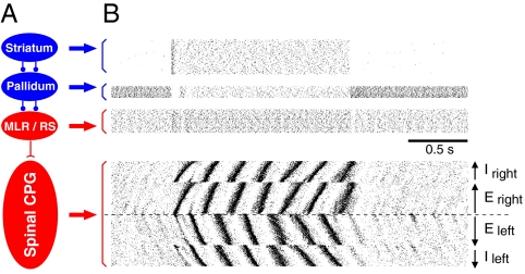
The vertebrate central nervous system is organized in modules that independently execute sophisticated tasks. Such modules are flexibly controlled and operate with a considerable degree of autonomy. One example is locomotion generated by spinal central pattern generator networks (CPGs) that shape the detailed motor output. The level of activity is controlled from brainstem locomotor command centers, which in turn, are under the control of the basal ganglia. By using a biophysically detailed, full-scale computational model of the lamprey CPG (10,000 neurons) and its brainstem/forebrain control, we demonstrate general control principles that can adapt the network to different demands. Forward or backward locomotion and steering can be flexibly controlled by local synaptic effects limited to only the very rostral part of the network. Variability in response properties within each neuronal population is an essential feature and assures a constant phase delay along the cord for different locomotor speeds.


Conflict of interest statement

The authors declare no conflict of interest.

Figures

Fig. 1.
Fig. 1.
Full-scale simulation of the spinal locomotor network of lamprey. (A) Mean firing rate (thick line) as a function of the somatic current injection. The shaded area shows maximum variation of the spike frequency in the simulated population. (B and C) Organization of the synaptic connections in transverse (B) and longitudinal (C) views of the spinal cord. Caudally directed projections dominate. E neurons project ipsilaterally, i.e., their axons do not cross the midline, and I neurons project contralaterally.
Fig. 2.
Fig. 2.
Control of the direction of swimming. (A) Pattern of neuron activity for the forward and backward swimming in specified segments on one side of the spinal cord. (B) Activity in four simulated populations, the left and right E and I interneurons. Dots correspond to the spikes in single neurons. Neurons are ordered in a rostrocaudal direction according to the arrows. (C and D) Controlling the intersegmental phase lag by the rostral command. Somatic current injection is applied to interneurons in the first 10 segments, with a linear decay (C Inset). The magnitude of the intersegmental phase lag varies continuously with the current injection for both the hemispinal cord (open squares) and intact spinal cord networks (filled circles; C). Only the left E population of the intact cord network is shown (D) in five different cases with different stimulation levels from +0.05 nA to −0.1 nA. In each sequence, the most rostral segment is represented in the uppermost position and the neurons in segment 100 in the lowermost position. Note that the positive phase lag is the largest with +0.05 nA applied to the 10 most rostral segments and the most negative with −0.1 nA.
Fig. 3.
Fig. 3.
Initiation of locomotor activity from the basal ganglia–brainstem. (A) Scheme of the basal ganglia–brainstem control of locomotor behavior. The neurons in the basal ganglia output stage, the pallidum, are during resting conditions tonically active, thus keeping motor centra in the brainstem (MLR, mesencephalic locomotor region and reticulospinal neurons, RS) under tonic inhibition. It is only after a strong excitatory input to the basal ganglia input stage, the striatum, that the striatal projections neurons can inhibit the tonic activity in the pallidal neurons, thus releasing the brainstem command centra from tonic inhibition. (B) This control of the spinal CPG is simulated. The resting CPG network receives a low synaptic drive from the brainstem motor centra (reticulospinal neurons, RS) because these are inhibited by pallidal cells. When the activity is suddenly released in the reticulospinal neurons because of disinhibition through the basal ganglia, the locomotor activity in the spinal network appears. Both an appropriate locomotor frequency and coordination is, as a consequence, immediately achieved.
Fig. 4.
Fig. 4.
Left–right steering of locomotion initiated from the basal ganglia. Locomotor activity was initiated by an enhanced activity in striatal neurons (blue trace) that, in turn, inhibits the tonic activity in pallidum (blue trace). The disinhibition of MLR/RS leads to an enhanced activity that turns on the spinal locomotor activity. Alternating activity is indicated by motoneuronal activity (MN) on the left (red) and right side (green). Activity of the neuronal pools is shown as relative changes of the firing rate in each population. Left and right MN activity is represented as the summed activity over the 10 most rostral segments. The drive signal to the striatal neurons is symmetric and bilateral and leads via the pallidum to a symmetric activation of the left and right MLR/RS populations. The model design for forward locomotion (blue trace) is identical to that in Fig. 3, but in addition, we have introduced two separate unilateral population of striatal neurons one for the left (red) and one for the right side (green), that will inhibit the corresponding unilateral subpopulations in the pallidum, which project specifically to the right or left side MLR/RS cells. The increased activity of the striatal right population (green) leads to the expected changes in the pallidal subpopulation and an elevation of the right MLR/RS population and finally an enhanced asymmetric MN alternations with the right side dominating. This would cause a turning to the right in the living animal. When the steering command is terminated, the network returns to the forward swimming mode.
Fig. 5.
Fig. 5.
Intersegmental coordination of neural activity. (A) Intensity of neural activity in a hemisegment, measured as subthreshold depolarization in motorneurons, for homogeneous network and a network with distributed parameters. (B) In the inhomogeneous network, the time delay d between the beginning of the depolarization and the crossing of the threshold for synaptic interaction between the segmental populations scales linearly with the cycle duration T. (C) Intersegmental phase lag as function of oscillation frequency. The relation for the homogeneous network is close to linear, y = kx (thick dashed line). The parameter variability in the inhomogeneous network changes this dependence to a near-constant relation, y = const (thick solid line). Shaded area shows the range of adaptation of the intersegmental delay.

References

    1. Grillner S, Hellgren J, Ménard A, Saitoh K, Wikström MA. Mechanisms for selection of basic motor programs–roles for the striatum and pallidum. Trends Neurosci. 2005;28:364–370. - PubMed
    1. Hikosaka O. GABAergic output of the basal ganglia. Prog Brain Res. 2007;160:209–226. - PubMed
    1. Kiehn O. Locomotor circuits in the mammalian spinal cord. Annu Rev Neurosci. 2006;29:279–306. - PubMed
    1. Feldman J, Del Negro CA. Looking for inspiration: New perspectives on respiratory rhythm. Nat Rev Neurosci. 2006;7:232–242. - PMC - PubMed
    1. Roberts A, Li WC, Soffe SR, Wolf E. Origin of excitatory drive to a spinal locomotor network. Brain Res Rev. 2008;57:22–28. - PubMed

Publication types