. 2009 Nov 10;106(45):19150-5.
doi: 10.1073/pnas.0909293106. Epub 2009 Nov 9.

Rescue of radiation-induced cognitive impairment through cranial transplantation of human embryonic stem cells

Affiliations

Rescue of radiation-induced cognitive impairment through cranial transplantation of human embryonic stem cells

Munjal M Acharya et al. Proc Natl Acad Sci U S A. .

Abstract

Cranial irradiation remains a frontline treatment for the control of tumor growth, and individuals surviving such treatments often manifest various degrees of cognitive dysfunction. Radiation-induced depletion of stem/precursor cell pools in the brain, particularly those residing in the neurogenic region of the hippocampus, is believed, in part, to be responsible for these often-unavoidable cognitive deficits. To explore the possibility of ameliorating radiation-induced cognitive impairment, athymic nude rats subjected to head only irradiation (10 Gy) were transplanted 2 days afterward with human embryonic stem cells (hESC) into the hippocampal formation and analyzed for stem cell survival, differentiation, and cognitive function. Animals receiving hESC transplantation exhibited superior performance on a hippocampal-dependent cognitive task 4 months postirradiation, compared to their irradiated surgical counterparts that did not receive hESCs. Significant stem cell survival was found at 1 and 4 months postirradiation, and transplanted cells showed robust migration to the subgranular zone throughout the dentate gyrus, exhibiting signs of neuron morphology within this neurogenic niche. These results demonstrate the capability to ameliorate radiation-induced normal tissue injury using hESCs, and suggest that such strategies may provide useful interventions for reducing the adverse effects of irradiation on cognition.


Conflict of interest statement

The authors declare no conflict of interest.

Figures
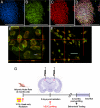
Fig. 1.
Fig. 1.
Pluripotency markers in hESCs. Immunocytochemical analysis of hESCs counter-stained with DAPI (A) shows strong expression and colocalization of the pluripotent markers Stage Specific Embryonic Antigen, (SSEA-4, B), and transcription factor Octamer-4, (Oct-4, C), shown as merged image (D). Colabeling of BrdU (red) and human specific nuclear antigen (green) in vitro (E) and in vivo (F) confirm the correspondence between BrdU labeled hESC and the presence of a specific human antigen. Orthogonal images (E and F) represent 3-D reconstructions of 1-μm confocal sections, and (F) were derived from a coronal section of a rat brain grafted with hESCs 4 months prior. (G) Schematic illustration of transplantation studies. Two-month-old athymic nude rats given 10 Gy head-only irradiation were grafted with hESC 2 days post-irradiation. At 4 months following these procedures, animals were subjected to cognitive testing then euthanized for the determination of transplanted cell survival and differentiation. A cohort of animals that did not undergo cognitive testing was also euthanized at 1 month post-transplant for survival and differentiation studies. (Scale bars, 200 μm in A–D; 2 μm in E; and 4 μm in F).
Fig. 2.
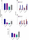
Fig. 2.
Human embryonic stem cell implantation improves radiation-induced impairments in novel place recognition (NPR). Rats were first familiarized with two identical objects in specific spatial locations in an open field, and total time spent exploring was assessed. Following a 5-min retention interval, they were re-presented with the same objects with one moved to a novel spatial location. (A) Irradiated animals (IRR) spent a significantly lower proportion of time exploring the novel place [ANOVA, P = 0.031, FPLSD, P < 0.015 vs. controls (Con) and vs. hESC-implanted controls (Con+hESC)]. In contrast, irradiated animals that received hESC injections (IRR+hESC) did not differ from either control group (P >0.25), and spent more time exploring the novel place than expected by chance (dashed line at 50%) although the latter was not found to be statistically significant (one sample t-test, P = 0.364). Nonetheless, the fact that the IRR+hESC group did not differ from controls suggests that performance was partially restored following hESC grafting. Twenty-four hours after the initial familiarization phase, rats were again presented with the same two objects, with one moved to a new spatial location. After the 24-h retention interval, (C) animals in the IRR group spent a significantly lower proportion of time exploring the novel place (ANOVA, P = 0.042, FPLSD, P = 0.007 vs. Con). In contrast, IRR+hESC animals did not differ from controls (P = 0.208). The results suggest that hESC transplantation partially rescued radiation-induced deficits on hippocampal dependent NPR task. Similar trends were observed when total time spent exploring the novel place was assessed after both the 5-min (B) and 24-h (D) retention intervals. Analysis of time spent exploring both objects during the initial familiarization phase revealed that IRR animals tended to spend less time engaged in exploration than both the Con and IRR+hESC group (E, ANOVA, P = 0.026, FPLSD, P = 0.068 vs. Con and P = 0.004 vs. IRR+hESC), which may partially explain why they showed impairments in NPR following the retention intervals. Data are presented as means + 1 S.E.M.
Fig. 3.
Fig. 3.
Survival and intrahippocampal location of transplanted hESCs. At 1 month postgrafting, hESCs are shown to incorporate extensively throughout the host-hippocampus (magnification, ×5–20 in A–C). Qualitatively similar patterns of migration were observed at 4 months post-grafting, albeit lower hESC numbers were evident (magnification, ×5–20 in D–F). Grafted cells were detected by BrdU immunostaining (dark brown nuclei) and counterstained with haematoxylin. Transplanted hESCs migrated extensively from the site of injection throughout the hippocampal formation (dentate gyrus, DG, dentate hilus, DH, granule cell layer, GCL, and CA1) and partially in the corpus callosum. Images shown were derived from irradiated animals. (Scale bars, 200 μm in A and D; 100 μm in B and E; and 50 μm in C and F.
Fig. 4.
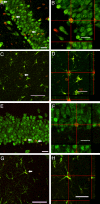
Fig. 4.
Differentiation of transplanted hESCs. At 1 month post-grafting (A–D), BrdU (red) positive hESCs were observed to differentiate into mature neurons (A and B) or astrocytes (C and D) as indicated by the colabeling of the neuron-specific nuclear antigen (NeuN, green) or glial fibrillary acidic protein (GFAP, green). Similar phenotypes were found 4 months post-grafting (E–H), where BrdU (red)-positive cells were also found to express markers of mature neurons (NeuN, E and F) and astrocytes (GFAP, G and H). Orthogonal reconstructions of BrdU+/NeuN+ colabeled cells (B and F) and BrdU+/GFAP+ colabeled cells (D and H) are shown at each post-grafting time. Images shown were derived from irradiated animals. (Scale bars, 20 μm in A and E; 50 μm in C and G; and 20 μm in B, D, F, H.)

References

    1. Walker MD, Strike TA, Sheline GE. An analysis of dose-effect relationship in the radiotherapy of malignant gliomas. Int J Radiat Oncol Biol Phys. 1979;5:1725–1731. - PubMed
    1. Sheline GE, Wara WM, Smith V. Therapeutic irradiation and brain injury. Int J Radiat Oncol Biol Phys. 1980;6:1215–1228. - PubMed
    1. Tofilon PJ, Fike JR. The radioresponse of the central nervous system: A dynamic process. Radiat Res. 2000;153:357–370. - PubMed
    1. Shaw E, et al. Single dose radiosurgical treatment of recurrent previously irradiated primary brain tumors and brain metastases: Final report of RTOG protocol 90–05. Int J Radiat Oncol Biol Phys. 2000;47:291–298. - PubMed
    1. Butler J, Rapp S, Shaw E. Managing the cognitive effects of brain tumor radiation therapy. Curr Treat Options Oncol. 2006;7:517–523. - PubMed

Publication types

Substances

LinkOut - more resources