. 2009 Nov 24;106(47):20021-6.
doi: 10.1073/pnas.0905686106. Epub 2009 Nov 9.

RAGE-mediated signaling contributes to intraneuronal transport of amyloid-beta and neuronal dysfunction

Affiliations

RAGE-mediated signaling contributes to intraneuronal transport of amyloid-beta and neuronal dysfunction

Kazuhiro Takuma et al. Proc Natl Acad Sci U S A. .

Abstract

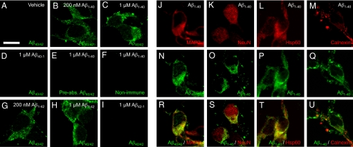
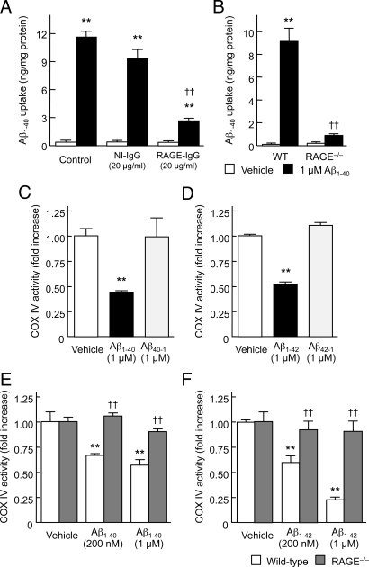
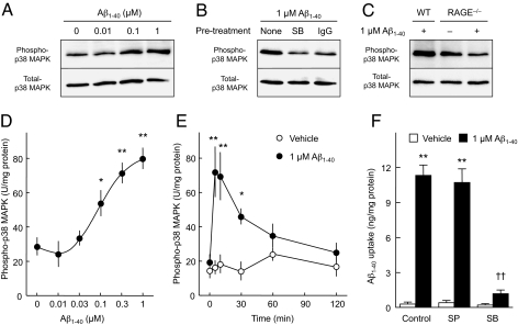
Intracellular amyloid-beta peptide (Abeta) has been implicated in neuronal death associated with Alzheimer's disease. Although Abeta is predominantly secreted into the extracellular space, mechanisms of Abeta transport at the level of the neuronal cell membrane remain to be fully elucidated. We demonstrate that receptor for advanced glycation end products (RAGE) contributes to transport of Abeta from the cell surface to the intracellular space. Mouse cortical neurons exposed to extracellular human Abeta subsequently showed detectable peptide intracellularly in the cytosol and mitochondria by confocal microscope and immunogold electron microscopy. Pretreatment of cultured neurons from wild-type mice with neutralizing antibody to RAGE, and neurons from RAGE knockout mice displayed decreased uptake of Abeta and protection from Abeta-mediated mitochondrial dysfunction. Abeta activated p38 MAPK, but not SAPK/JNK, and then stimulated intracellular uptake of Abeta-RAGE complex. Similar intraneuronal co-localization of Abeta and RAGE was observed in the hippocampus of transgenic mice overexpressing mutant amyloid precursor protein. These findings indicate that RAGE contributes to mechanisms involved in the translocation of Abeta from the extracellular to the intracellular space, thereby enhancing Abeta cytotoxicity.


Conflict of interest statement

Conflict of interest statement: D. Stern is a consultant for TransTech Pharma.

Figures

Fig. 1.
Fig. 1.
Confocal images of Aβ, MAP2, NeuN, Hsp60, and calnexin in cortical neurons after exposure to Aβ-related peptides. Cells were exposed to the indicated concentration [or 1μM (JU)] of human Aβ1−40, Aβ1−42, reversed Aβ (Aβ40−1 and Aβ42−1), or vehicle for 60 min, fixed in 3% PFA, and stained by [anti-human Aβ (clone 4G8) (A–D, GI, N, and R), anti-Aβ1–40 (O–P and S–U), preabsorbed anti-Aβ (clone 4G8) (E) or non-immune IgG (F)]/Alexa Fluor 488 anti-IgG (green), anti-MAP2/Alexa Fluor 568 anti-IgG (red) (J and R) and [anti-NeuN (K and S), anti-Hsp 60 (L and T), or anti-calnexin (M and U)]/Alexa Fluor 546 anti-IgG (red). Scale bar, 10 μm. Hoechst 33342 staining and phase contrast images of the same field of cells in panels of A, C, H, D, or Fig. S4 A–D are represented in Fig. S4 E–H and M–P, respectively.
Fig. 2.
Fig. 2.
Immunoelectron microscopy of Aβ in cortical neurons after exposure to Aβ. Cells were prepared from wild-type (WT) (A and B) and RAGE−/− mice (C), exposed to human 1 μM Aβ1–42 for 60 min, fixed in 4% PFA and 0.1% glutaraldehyde, and the ultra-thin sections were stained with rabbit anti-Aβ1–42/donkey anti-rabbit IgG conjugated to colloidal gold (18 nm particle). Arrows denote mitochondria. (Scale bar, 200 nm.) Two negative controls, in which cells were treated with vehicle or stained with non-immune IgG (NI-IgG), are represented in Fig. S5. (D) Quantification of Aβ immunogold particles in WT and RAGE−/− neurons after exposure to Aβ. Numbers of gold particles were counted per field of each microscopic image including two negative controls and expressed as mean ± SEM; ***, P < 0.001, versus WT; Unpaired t-test.
Fig. 3.
Fig. 3.
Blocking RAGE or genetic deletion of the receptor suppresses Aβ uptake and minimizes Aβ-induced mitochondrial dysfunction in cortical neurons. Intracellular levels of human Aβ1–40 (A and B) and COX IV activity (C–F) were assayed 60 min (A and B) and 24 h (C–F) after exposure to the indicated Aβ peptides. (A) Effect of a neutralizing antibody to RAGE. Cells were pre-treated with 20 μg/mL of anti-RAGE (N-16) IgG or NI-IgG for 2 h, and then exposed to 1 μM human Aβ1–40. (B, E, and F) Effect of genetic deletion of RAGE. Cells prepared from WT or RAGE−/− mice were exposed to the indicated concentrations of human Aβ1–40 (B and E) or Aβ1–42 (F). (C and D) Aβ-related peptides with the reverse sequence have no effect on mitochondrial function in cortical neurons. Cells prepared from wild-type mice were exposed to 1 μM human Aβ1–40 or Aβ40–1 (C), and 1 μM human Aβ1–42 or Aβ42–1 (D). Data represent mean ± SEM; **, P < 0.01, versus vehicle- and reversed Aβ-treated cells (A–D), or Aβ-treated RAGE−/− neurons (E and F); ††, P < 0.01, versus control (A and B) or WT (E and F).
Fig. 4.
Fig. 4.
Aβ-stimulated p38 MAPK activation is required for Aβ uptake in cortical neurons. (A–C) Immunoblot analyses of phospho-p38 MAPK in cortical neurons treated with Aβ1–40. Cells were exposed to the indicated concentrations of human Aβ1–40 for 10 min, lysed, and subjected to SDS/PAGE. Typical immunoblot images detected by antibodies against phospho-p38 MAPK (A–C, upper) and total-p38 MAPK (A–C, lower) are shown from 3–6 independent experiments. (A) Dose-dependency. (B) The p38 MAPK inhibitor SB 203580 (1 μM; SB) and anti-RAGE (N-16) IgG (20 μg/mL; IgG) were added 30 min and 2 h before exposure to Aβ1–40, respectively. (C) Phospho-p38 MAPK levels in neurons from RAGE−/− mice after exposure to Aβ1–40. WT, wild-type. (D and E) Phospho-p38 MAPK levels were determined by ELISA. (D) Dose-dependency. Cells were exposed to the indicated concentration of human Aβ1–40 for 10 min. (E) Time course. Cortical neurons were exposed to 1 μM of human Aβ1–40 for the indicated time. (G) Effects of JNK and p38 MAPK inhibitors on intracellular levels of human Aβ1–40 in cultured neurons exposed to Aβ. Cells were pretreated with the JNK inhibitor SP 600125 (SP, 20 μM), or SB 203580 (SB, 1 μM), for 30 min, and then exposed to 1 μM human Aβ1–40 for 60 min. Intracellular Aβ1–40 concentrations were determined by ELISA. Data represent mean ± SEM; *, P < 0.05, **, P < 0.01, versus none (0 μM Aβ1–40) (D and F) and 0-time (E); ††, P < 0.01, versus control (no inhibitor).

References

    1. Yankner BA. Mechanisms of neuronal degeneration in Alzheimer's disease. Neuron. 1996;16:921–932. - PubMed
    1. LaFerla FM, Oddo S. Alzheimer's disease: Aβ, tau, and synaptic dysfunction. Trends Mol Med. 2005;11:170–176. - PubMed
    1. Gouras GK, et al. Intraneuronal Aβ42 accumulation in human brain. Am J Pathol. 2000;156:15–20. - PMC - PubMed
    1. Takahashi RH, et al. Intraneuronal Alzheimer Aβ42 accumulates in multivesicular bodies and is associated with synaptic pathology. Am J Pathol. 2002;161:869–879. - PMC - PubMed
    1. LaFerla FM, Green KN, Oddo S. Intracellular amyloid-β in Alzheimer's disease. Nat Rev Neurosci. 2007;8:499–509. - PubMed

Publication types

MeSH terms

Substances