. 2010 Oct;43(4):422-31.
doi: 10.1165/rcmb.2009-0224OC. Epub 2009 Nov 9.

Plasmacytoid dendritic cells prevent cigarette smoke and Chlamydophila pneumoniae-induced Th2 inflammatory responses

Affiliations

Plasmacytoid dendritic cells prevent cigarette smoke and Chlamydophila pneumoniae-induced Th2 inflammatory responses

Rosalinda Sorrentino et al. Am J Respir Cell Mol Biol. 2010 Oct.

Abstract

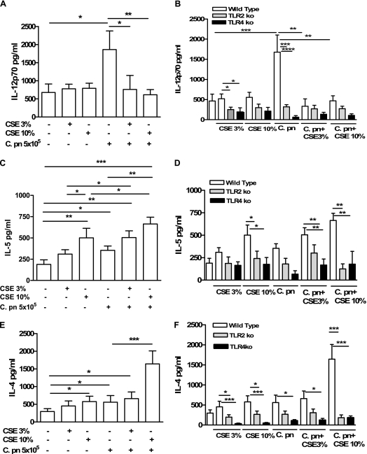
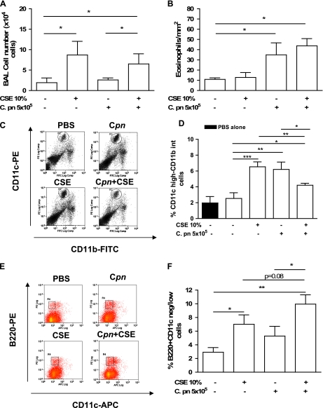
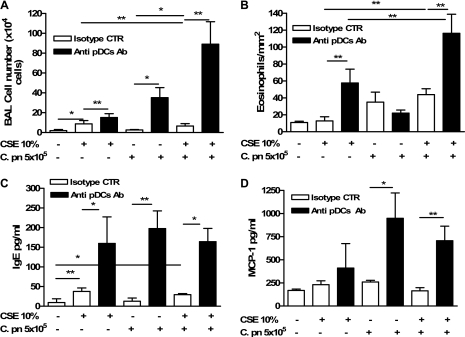
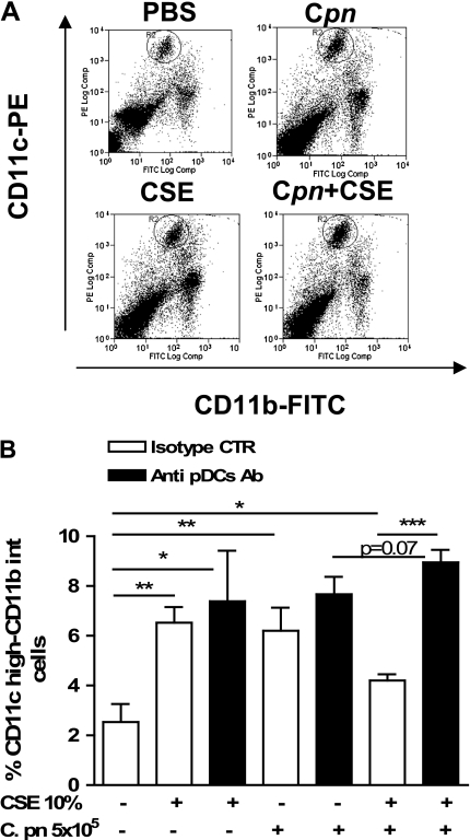
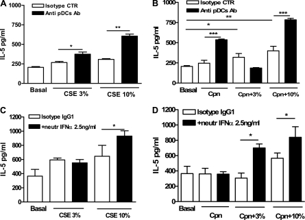
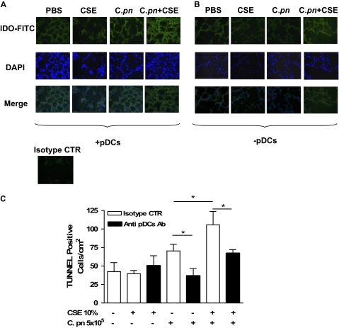
Smoking promotes the development of allergic asthma and pneumonia. Chlamydophila pneumoniae lung infection is associated with an increased risk for asthma, inducing an immune response regulated by dendritic cells (DCs). This study sought to determine whether exposure to cigarette smoke modulates the functional activity of CD11c-positive DCs in the lung, with and without concomitant C. pneumoniae infection. Bone marrow-derived DCs (BMDCs) were exposed in vitro to cigarette smoke extract (CSE) and/or live C. pneumoniae (Cpn), and then adoptively transferred intratracheally into wild-type mice. Although CSE plus Cpn appeared to exert an additive effect on the production of Th2 cytokines in vitro, we did not see this effect in vivo. However, the adoptive transfer of DCs pulsed with both CSE and C. pneumoniae into the lungs of naive mice led to an influx of plasmacytoid DCs (pDCs) that suppressed the Th2 skewing ability of the transferred BMDCs. The depletion of pDCs by antibody restored the Th2 skewing ability of the BMDCs. The expression of indoleamine-2,3-dioxygenase in the lung was reduced after the depletion of pDCs, and blocking IFN-α in vitro prevented the ability of pDCs to inhibit the Th2 responses induced by myeloid DCs (mDCs), suggesting their potential involvement in the mechanism of altered polarization. In conclusion, exposure to cigarette smoke skews C. pneumoniae-induced mDCs responses toward a Th2 bias in the lung, which is prevented by pDCs. We propose that pDCs may play a major role in the immunosuppressive lung environment in smokers with C. pneumoniae infection.


Figures

Figure 1.
Figure 1.
The cigarette smoke extract (CSE) skews the C. pneumoniae–induced Th1-like response toward a Th2-like response in vitro. The bone marrow–derived dendritic cells (BMDCs) were treated with CSE alone (3% and 10%) and/or C. pneumoniae, and 24 hours later the supernatants were harvested. Concentrations of IL-12p70 (A, B), IL-5 (C, D), and IL-4 (E, F) were measured using ELISA. (A, C, E) Wild-type BMDC mice (open bars). (B, D, F) Wild-type (open bars), TLR2 KO (shaded bars), and TLR4 KO (solid bars) mice. There were 12 experiments, and each treatment was performed in triplicate. Statistically significant differences are denoted as *P < 0.05, **P < 0.01, and ***P < 0.0001, as determined by one-way ANOVA and the Student t test.
Figure 2.
Figure 2.
The adoptive transfer of CSE-pulsed and/or C. pneumoniae–pulsed BMDCs modulates the immune response in the lung. The BMDCs were pulsed ex vivo overnight with PBS, CSE (10%), C. pneumoniae (5 × 10⁵ IFUs/ml), or both, and then adoptively transferred (intratracheally; 5 × 10⁵ cells/mouse) into wild-type recipient mice. Mice were killed 5 days later, and lungs were excised and properly stained for CD11c-PE and CD11b-FITC. (A) BAL cell numbers. (B) Eosinophil numbers. (C, D) Lung mDCs were expressed as percentages of CD11c highly positive and CD11b intermediately positive cells. (E, F) In the same manner, lung pDCs were determined as CD11c negative/low and B220 high. (D) Solid bar represents values of mDCs in the lungs of wild-type mice injected with 100 μl of PBS. (C, E) Experiments representative of bar graphs in D and F, respectively. Data represent mean ± SEM, n = 9/each group. Experiments were performed on 2 different days. Statistically significant differences are denoted as *P < 0.05, **P < 0.01, and ***P < 0.005, as determined by one-way ANOVA and the Student t test.
Figure 3.
Figure 3.
Depletion of plasmacytoid dendritic cells (pDCs) results in increased Th2 inflammation after transfer of BMDCs pulsed with CSE, C. pneumoniae, or both. Mice were injected intravenously with either isotype control Ab (IgG2a, open bars), or with 120G8 Ab (solid bars), that is, the specific Ab for the depletion of pDCs. The intravenous injection of IgG2a or 120G8 Ab was performed 3 days after the adoptive transfer of CSE-pulsed and/or C. pneumoniae–pulsed BMDCs. The bronchoalveolar lavage (BAL) cell numbers (A) and eosinophil numbers (B) were evaluated as described in the text. The ELISA experiments were performed to evaluate IgE (C) and MCP-1 (D) in BAL-derived supernatants. Data represent mean ± SEM; n = 9 and n = 6 for pDC depletion experiments. Two separate experiments were performed. Statistically significant differences are denoted as *P < 0.05 and **P < 0.01, as determined by one-way ANOVA and the Student t test.
Figure 4.
Figure 4.
Depletion of pDCs results in increased mDCs in the lung. The BMDCs were treated overnight with PBS, CSE (10%), C. pneumoniae (5 × 10⁵ IFUs/ml), or both, and then adoptively transferred (intratracheally; 5 × 10⁵ cells/mouse) into wild-type recipient mice. Mice were killed 5 days later, and lungs were excised and properly stained for CD11c-PE and CD11b-FITC. (A, B) Lung mDCs were expressed as percentages of CD11c highly positive CD11b intermediately positive cells. (A) An experiment representative of bar graph in B. Data represent mean ± SEM, n = 9 per group. Experiments were performed on 2 different days. Statistically significant differences are denoted as *P < 0.05, **P < 0.01, and ***P < 0.0001, as determined by one-way ANOVA and the Student t test.
Figure 5.
Figure 5.
Lung CD11c+ cell release increased IL-5 ex vivo when pDCs were depleted before purification. The BMDCs were treated overnight with PBS, CSE (10%), C. pneumoniae (5 × 10⁵ IFUs/ml) or both, and then adoptively transferred (intratracheally; 5 × 10⁵ cells/mouse) into wild-type recipient mice. Two days later, 120G8 Ab or isotype control were intravenously injected (150μg/ml) lung CD11c+ cells were isolated by means of a technique using Miltenyi Biotec microbeads 3 days later. (A, B) IL-5 concentrations were measured using ELISA. Neutralizing Ab for IFN-α (2.5 ng/ml) or isotype control was added to CD11c+ cells isolated from the lung, and IL-5 concentrations were measured when exposed to CSE and C. pneumoniae (C, D). Concentrations of IL-5 were analyzed using ELISA in cell-free supernatants. Data represent mean ± SEM, n = 9 and n = 6 for pDC depletion experiments. Experiments were performed on 2 different days. Statistically significant differences are denoted as *P < 0.05, **P < 0.01, and ***P < 0.0001, as determined by one-way ANOVA and the Student t test.
Figure 6.
Figure 6.
The BMpDCs expressed higher levels of IFN-α and IDO in response to CSE and/or C. pneumoniae stimulation. (A) The BMpDCs were treated with CSE and/or C. pneumoniae, and concentrations of IFN-α mRNA were measured using real-time PCR. (B) Concentrations of IDO protein were evaluated by means of immunoblotting. Data are expressed as ratios between IFN-α (A) or IDO (B) compared with GAPDH expression. CpG oligodeoxynucleotides (1 μg/ml) was used as positive control for IFN-α mRNA expression. Quantitative data were obtained by analyzing optical density values with ImageJ software. Data represent mean ± SEM, n = 3 (A) and n = 3 (B). Statistically significant differences are denoted as *P < 0.05, and **P < 0.005, as determined by one-way ANOVA and the Student t test.
Figure 7.
Figure 7.
The depletion of pDCs significantly decreased IDO and apoptotic cells in the lung. Mice were adoptively transferred with CSE-pulsed and/or C. pneumoniae–pulsed BMDCs, and lung sections were stained for IDO expression and TUNEL assay. The expression of IDO was determined by means of an immunofluorescence technique, using FITC either in the presence (A) or after the depletion (B) of pDCs. The isotype control (rat IgG2b) was used as negative control. (C) A TUNEL assay was performed, and positive cells were quantified. Data represent mean ± SEM, and each experiment was performed at least five times for each type of experimental condition. Statistically significant differences are denoted as *P < 0.05, as determined by one-way ANOVA and the Student t test.

Similar articles

Cited by

References

    1. Chalmers GW, MacLeod KJ, Thomson L, Little SA, McSharry C, Thomson NC. Smoking and airway inflammation in patients with mild asthma. Chest 2001;120:1917–1922. - PubMed
    1. Hylkema MN, Sterk PJ, de Boer WI, Postma DS. Tobacco use in relation to COPD and asthma. Eur Respir J 2007;29:438–445. - PubMed
    1. D'Hulst AI, Maes T, Bracke KR, Demedts IK, Tournoy KG, Joos GF, Brusselle GG. Cigarette smoke-induced pulmonary emphysema in scid-mice. Is the acquired immune system required? Respir Res 2005;6:147. - PMC - PubMed
    1. Reibman J, Hsu Y, Chen LC, Bleck B, Gordon T. Airway epithelial cells release MIP-3α/CCL-20 in response to cytokines and ambient particulate matter. Am J Respir Cell Mol Biol 2003;28:648–654. - PubMed
    1. Naiki Y, Sorrentino R, Wong MH, Michelsen KS, Shimada K, Chen S, Yilmaz A, Slepenkin A, Schröder NW, Crother TR, et al. TLR/MyD88 and liver X receptor alpha signaling pathways reciprocally control Chlamydia pneumoniae-induced acceleration of atherosclerosis. J Immunol 2008;181:7176–7185. - PMC - PubMed

Publication types

MeSH terms