

Intraoperative evaluation of the spiral nerve cuff electrode on the femoral nerve trunk
- PMID: 19901448
- PMCID: PMC2927973
- DOI: 10.1088/1741-2560/6/6/066005
Intraoperative evaluation of the spiral nerve cuff electrode on the femoral nerve trunk
Abstract
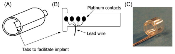
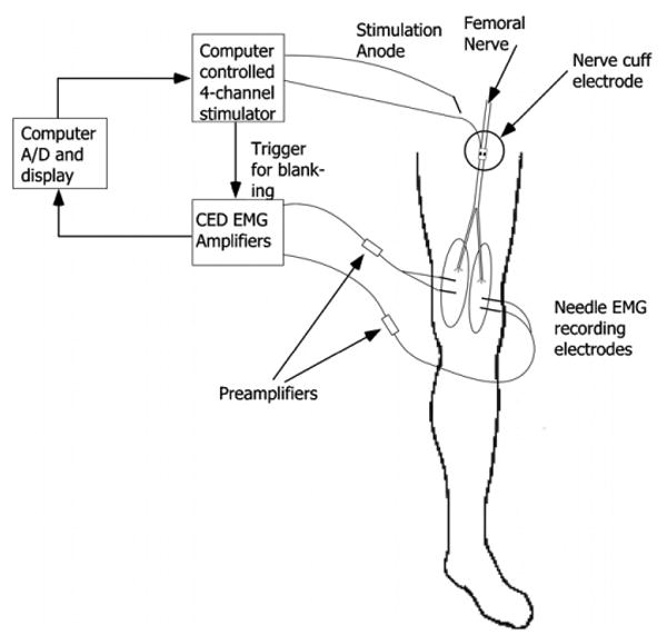
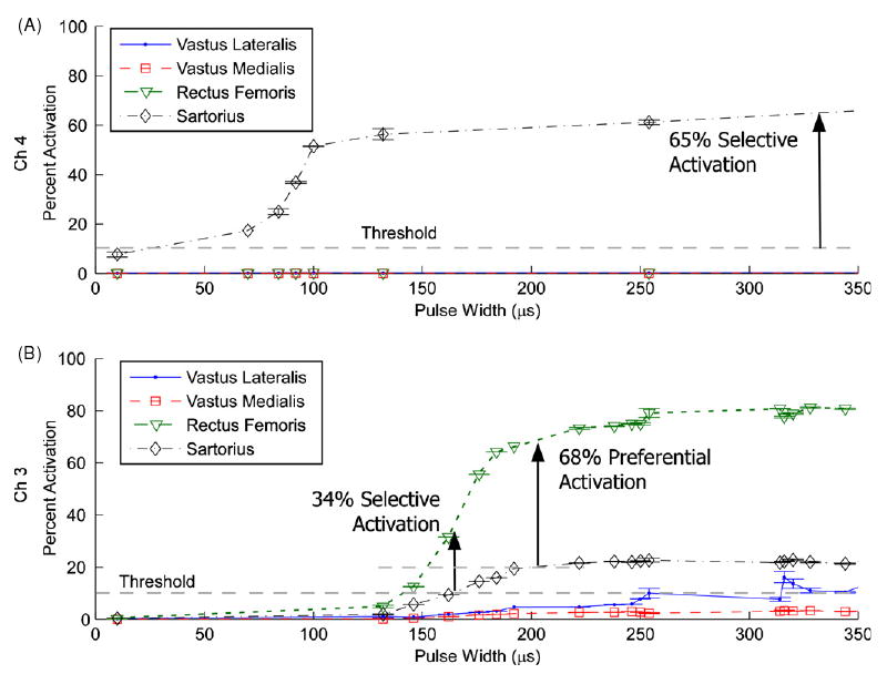
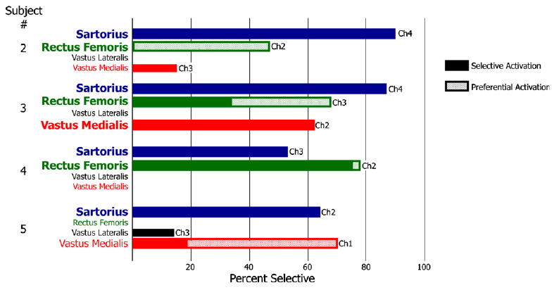
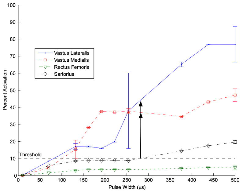
Evaluation of the Case Western Reserve University spiral nerve cuff electrode on the femoral nerve trunk was performed intraoperatively in four subjects undergoing femoral-popliteal bypass surgery. The threshold, nerve size and selective activation capabilities of the electrode were examined. The activation thresholds for the first muscle to be recruited were 6.3, 9, 10.6, and 37.4 nC with pulse amplitudes ranging from 0.3 to 1 mA. The femoral nerve was found to have an elliptical cross-section with a major axis average length of 9 mm (8-12 mm) and a minor axis length of 1.5 mm. In all four subjects selective activation of the sartorius was obtained. In two subjects, the rectus femoris could also be selectively activated and in one subject the vastus medialis was selectively activated. Each electrode had four independent contacts that were evaluated separately. Small air bubbles were formed in the space over some contacts, preventing stimulation. This occurred in one contact in each electrode, leaving three effective stimulation channels. This issue has been corrected for future studies.
Figures
References
-
- Memberg WD, Peckham PH, Keith MW. A surgically-implanted intramuscular electrode for an implantable neuromuscular stimulation system. IEEE Trans Rehabil Eng. 1994;2:80–91.
-
- Akers JM, Peckham PH, Keith MW, Merritt K. Tissue response to chronically stimulated implanted epimysial and intramuscular electrodes. IEEE Trans Rehabil Eng. 1997;5:207–20. - PubMed
-
- Uhlir JP, Triolo RJ, Davis JA, Jr, Bieri C. Performance of epimysial stimulating electrodes in the lower extremities of individuals with spinal cord injury. IEEE Trans Neural Syst Rehabil Eng. 2004;12:279–87. - PubMed
-
- Naples GG, Mortimer JT, Scheiner A, Sweeney JD. A spiral nerve cuff electrode for peripheral nerve stimulation. IEEE Trans Biomed Eng. 1988;35:905–16. - PubMed
Publication types
MeSH terms
Grants and funding
LinkOut - more resources
Full Text Sources
Other Literature Sources