. 2009 Nov;3(6):448-61.
doi: 10.4161/chan.3.6.10216. Epub 2009 Nov 30.

A novel N-terminal motif of dipeptidyl peptidase-like proteins produces rapid inactivation of KV4.2 channels by a pore-blocking mechanism

Affiliations

A novel N-terminal motif of dipeptidyl peptidase-like proteins produces rapid inactivation of KV4.2 channels by a pore-blocking mechanism

Henry H Jerng et al. Channels (Austin). 2009 Nov.

Abstract

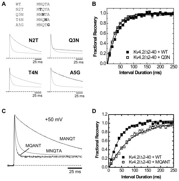
The somatodendritic subthreshold A-type K(+) current in neurons (I(SA)) depends on its kinetic and voltage-dependent properties to regulate membrane excitability, action potential repetitive firing, and signal integration. Key functional properties of the K(V)4 channel complex underlying I(SA) are determined by dipeptidyl peptidase-like proteins known as dipeptidyl peptidase 6 (DPP6) and dipeptidyl peptidase 10 (DPP10). Among the multiple known DPP10 isoforms with alternative N-terminal sequences, DPP10a confers exceptionally fast inactivation to K(V)4.2 channels. To elucidate the molecular basis of this fast inactivation, we investigated the structure-function relationship of the DPP10a N-terminal region and its interaction with the K(V)4.2 channel. Here, we show that DPP10a shares a conserved N-terminal sequence (MNQTA) with DPP6a (aka DPP6-E), which also induces fast inactivation. Deletion of the NQTA sequence in DPP10a eliminates this dramatic fast inactivation, and perfusion of MNQTA peptide to the cytoplasmic face of inside-out patches inhibits the K(V)4.2 current. DPP10a-induced fast inactivation exhibits competitive interactions with internally applied tetraethylammonium (TEA), and elevating the external K(+) concentration accelerates recovery from DPP10a-mediated fast inactivation. These results suggest that fast inactivation induced by DPP10a or DPP6a is mediated by a common N-terminal inactivation motif via a pore-blocking mechanism. This mechanism may offer an attractive target for novel pharmacological interventions directed at impairing I(SA) inactivation and reducing neuronal excitability.


Figures

Figure 1
Figure 1. The Most Upstream of the Alternatively Spliced First Exons of DPP10a and DPP6a (Exon 1a) Encode Highly Similar Amino Acid Sequences
(A) Alignment of amino acids sequences encoded by Exon 1a of DPP10 and DPP6 from various species. Sequences were obtained by mining cDNA and genomic databases. Identical residues between DPP10a and DPP6a are shown in bold; consensus residues, underlined. Notice the identity between residues 1-5 (MNQTA) of these DPLP variants. (B) Genomic organization of rat DPP10 and DPP6 genes, including Exon 1’s of known splice variants (DPP10: DPP10a, DPP10c, DPP10d; DPP6: DPP6a, DPP6-K, DPP6-L, DPP6-S). DPP10 rat gene is located on chromosome 13; DPP6, chromosome 4.
Figure 2
Figure 2. DPP10a and DPP6a Introduce a Similar Rapid Phase of Inactivation in Kv4.2+KChIP3a Channels
(A) DPP6a produces kinetic effects similar to those of DPP10a and very different from those of DPP6-S. Families of current traces were elicited by 10-mV step depolarizations of 1- or 5-sec duration from −100 mV to +60 mV, starting from a holding potential of −100 mV. Only the first 350 ms are shown. Inactivation kinetics at +50 mV were measured using multi-exponential fits, and based on the residuals (actual current traces minus fitted traces) Kv4.2+KChIP3a plus DPP10a or DPP6a requires three time constants to properly describe the decay (τ0, τ1, and τ2). However, the inactivation of Kv4.2+KChIP3a+DPP6-S channel is well-described by two time constants. Based on similarity of time constant values to currents modified by a-variants, the time constants were named τ1 and τ2. (B) The time course of inactivation at +50 mV. Notice the match between the fast time constants of Kv4.2+KChIP3a+DPP10a and Kv4.2+KChIP3a+DPP6a. (C) The fractional amplitudes of the inactivating components at +50 mV. The majority of the current undergoes fast inactivation in the presence of the intact DPP10a and DPP6a N-terminal domains. (D) Recovery from inactivation of Kv4.2+KChIP3a coexpressed with DPP6-S, DPP6a, and DPP10a at −100 mV. The standard two-pulse protocol was used, with a 1-sec long prepulse to +50 mV. (E) The peak conductance-voltage (Gp-V) relationships and steady-state inactivation curves for the channel complexes under study. Gp-V is best fit with a 4th-powered Boltzmann. Steady-state inactivation was fit with a single Boltzmann function. The symbols used are the same as in Fig. 2D, with I/Imax and Gp/Gp-max data points represented by open and closed symbols, respectively. The Gp-V and steady-state inactivation curves are quite similar, despite differing inactivation kinetics. Asterisks indicate statistical significance with p < 0.05 when compared to corresponding control values.
Figure 3
Figure 3. Deletion of DPP10a AA2-5 (NQTA) Is Sufficient to Eliminate DPP10a-mediated Fast Component of Inactivation
(A, B) Representative normalized current traces at +50 mV for Kv4.2/Δ2-40 coexpressed with DPP10a/Δ2-20 (A) and DPP10a/ΔNQTA (B). Traces were overlapped with normalized Kv4.2/Δ2-40+DPP10a trace (thin lines) to show kinetic differences. Rapid inactivation by DPP10a is eliminated by these N-terminal deletions. In panel B inset, traces are shown in smaller time scale to show similar activation kinetics. (C) Time constants of inactivating components at +50 mV for the channels under study. (D) Fractional amplitudes for inactivating components at +50 mV. (E) Normalized peak conductance-voltage (Gp-V) relationships. The Gp-V curves were analyzed as described in Materials and Methods. Deletions of MNQTA and residues 2-20 similarly produce ~ 15 mV leftward shift in the V0.5 value. (F) Measurements of steady-state inactivation at indicated membrane potentials. According to Boltzmann fit, no significant changes in midpoint or slope were produced by the deletions. Asterisks indicate statistical significance with p < 0.05 when compared to corresponding control values.
Figure 4
Figure 4. Deletion of the NQTA Residues Also Eliminates DPP10a-mediated Fast Inactivation in Cultured Mammalian Cells
(A) Cell-attached patch recording of Kv4.2/Δ2-40+DPP10a channels expressed in tsA-201 cells. The protocol for step depolarizations is indicated by the voltage trace inset. DPP10a-mediated fast inactivation is similar between tsA-201 cells and oocytes. (B) Deletion of NQTA residues leads to loss of DPP10a-mediated fast inactivation and the observed dramatic slowing of current decay. (C) Normalized current traces at +92 mV illustrate the dramatic slowing of inactivation due to DPP10a N-terminal truncation. Inset shows no change in activation kinetics, indicating that NQTA deletion does not affect Kv4.2-DPP10 association.
Figure 5
Figure 5. DPP10a-mediated Fast Inactivation is Sensitive to Conservative Substitutions and Sequence Rearrangement of the MNQTA Motif
(A) Normalized whole-ooyte currents at +50 mV from Kv4.2/Δ2-40 channels co-expressed with the indicated DPP10a point mutant. The traces were overlapped with that of normalized Kv4.2/Δ2-40+DPP10a for comparison. (B) The effect of Q3N mutation on the kinetics of recovery from inactivation at −100 mV. The duration of the control pulse was set at 25 ms to allow measurement of recovery from fast inactivation. Lines through the points represent the best fit, assuming a mono-exponential rise. (C) Normalized traces at +50 mV for Kv4.2/Δ2-40 co-expressed with either wild-type DPP10a, the MQANT mutant, or the MANQT mutant. (D) The time course of recovery from inactivation at −100 mV for channels co-expressed with wild-type DPP10a or the MQANT mutant.
Figure 6
Figure 6. Proportional Inhibition of Peak Current and Slowing of Inactivation of Kv4.2+DPP10a Channels by TEA Suggest Competition between Internal TEA and DPP10a-mediated Inactivation
(A) 10 mM internal TEA reduces Kv4.2/Δ2-40+DPP10a peak current by ~ 46%. Current traces were fitted with 2-exponential functions, and the dashed traces show the decay of the fast component before (black) and after (red) TEA application. Crossing-over of traces illustrates the slowdown of inactivation by internal TEA. (B) TEA-induced slowdown of inactivation, compared by overlapped normalized traces. Both the current inhibition and slowing of inactivation are reversed by washout. (C) Proportionality of fold inhibition and fold inactivation slowing by internal TEA. TEA (5 and 10 mM) was tested for potency in current inhibition and slowing. A direct proportionality is indicated by the identity line drawn, which indicates a 1:1 ratio between fold slowing and fold inhibition. Numbers indicate TEA concentration.
Figure 7
Figure 7. MNQTA Peptide Reversibly Inhibits Kv4.2/Δ2-40 Channel Associated with DPP10a/ΔNQTA
(A) Outward currents at +42 mV mediated by Kv4.2/Δ2-40 channels co-expressed with DPP10a/ΔNQTA. Traces show the reversible suppression of peak current by 300 μM MNQTA peptide application. (B) Timeline of a representative experiment showing the effect of internal perfusion of MNQTA peptide and TEA. Shaded bars indicate the duration of the applications.
Figure 8
Figure 8. External K+ Does Not Affect DPP10a-mediated Fast Inactivation
Outward currents at +50 mV from oocytes injected with cRNAs for (A) Kv4.2/Δ2-40, (B) Kv4.2/Δ2-40 and DPP10a, and (C) Kv4.2/Δ2-40 and DPP10a/ΔNQTA. Pulses were repeated after external K+ concentration was increased from 2 mM to 98 mM by replacing equivalent molar concentrations of Na+. Note that the fast component of inactivation for Fig. 7B is not affected by elevated K+, whereas the slow components are dramatically different. Inactivation of currents in Figs. 7A and 7C are markedly accelerated by the ion manipulation. (D) Quantitation of time constants for inactivation at +50 mV for the channels under study. (E) Quantitation of fractional amplitudes for inactivating components at +50 mV. Red bars and traces represent results from 98 mM external K+; black bars and traces, 2 mM external K+. Asterisks indicate statistical significance for the value under elevated [K+]ext with p < 0.05.
Figure 9
Figure 9. Elevated External K+ Concentration Accelerates Recovery from MNQTA-dependent Fast Inactivation
(A) Representative traces of Kv4.2/Δ2-40+DPP10a currents elicited by a two-pulse protocol with external K+ at 2 (black) or 98 (red) mM concentrations. After inactivating channels with +50 mV depolarizing pulse for 25 ms, recovery at −100 mV membrane potential was checked by a second pulse to +50 mV. Peak current returns more rapidly in elevated [K+}ext. (B) Time course of recovery from inactivation is significantly faster in 98 mM than in 2 mM [K+]ext. The lines represent single exponential fits to the data. (C) The kinetics for recovery from inactivation matches that of channel closing. A representative trace for a two-pulse experiment in 98 mM [K+]ext is overlapped with the data points of the peak recovered current, before (closed red symbols) and after the values were vertically offset and normalized to the tail current. Amplitude of the tail current at the end of the first pulse was determined by extrapolations of monoexponential fits. (D) Recovery from inactivation of Kv4.2/Δ2-40 channels coexpressed with DPP10a/ΔNQTA is unaffected by increasing [K+]ext.
Figure 10
Figure 10. Kv4 inactivation gating and open-state block by MNQTA ID and TEA
Simplified state diagram showing the closed (C), open (O), and closed-inactivated (IC) states. We hypothesize that during a prolonged depolarization, Kv4 channels coexpressed with DPP10a or DPP6a open and are transiently blocked by the MNQTA peptide (BMNQTA). The internal TEA competes with MNQTA peptide for the open channel by forming the BTEA state, with rapid on- and off-rates. The occupancy of either BMNQTA or BTEA states impairs the ability of Kv4 channels to close and ultimately accumulate in the IC state, thought to be the absorbing inactivated state of Kv4 channels., Pairs of arrows represents transitions between states with their respective forward and reverse rates. Dashed arrows indicate transitions to closed and closed-inactivated states relatively distant from the open states, which were removed for simplification.

Similar articles

Cited by

References

    1. Jerng HH, Pfaffinger PJ, Covarrubias M. Molecular physiology and modulation of somatodendritic A-type potassium channels. Mol Cell Neurosci. 2004;27:343–369. - PubMed
    1. Birnbaum SG, Varga AW, Yuan LL, Anderson AE, Sweatt JD, Schrader LA. Structure and function of Kv4-family transient potassium channels. Physiol Rev. 2004;84:803–833. - PubMed
    1. Covarrubias M, Bhattacharji A, De Santiago-Castillo JA, Dougherty K, Kaulin YA, Na-Phuket TR, Wang G. The neuronal Kv4 channel complex. Neurochem Res. 2008;33:1558–1567. - PMC - PubMed
    1. Maffie J, Rudy B. Weighing the evidence for a ternary protein complex mediating A-type K+ currents in neurons. J Physiol. 2008;586:5609–5623. - PMC - PubMed
    1. An WF, Bowlby MR, Betty M, Cao J, Ling HP, Mendoza G, Hinson JW, Mattsson KI, Strassle BW, Trimmer JS, Rhodes KJ. Modulation of A-type potassium channels by a family of calcium sensors. Nature. 2000;403:553–556. - PubMed

Publication types

Substances

LinkOut - more resources