. 2009 Nov 10;4(11):e7615.
doi: 10.1371/journal.pone.0007615.

Phenylalanine-rich peptides potently bind ESAT6, a virulence determinant of Mycobacterium tuberculosis, and concurrently affect the pathogen's growth

Affiliations

Phenylalanine-rich peptides potently bind ESAT6, a virulence determinant of Mycobacterium tuberculosis, and concurrently affect the pathogen's growth

Krishan Kumar et al. PLoS One. .

Abstract

Background: The secretory proteins of Mycobacterium tuberculosis (M. tuberculosis) have been known to be involved in the virulence, pathogenesis as well as proliferation of the pathogen. Among this set, many proteins have been hypothesized to play a critical role at the genesis of the onset of infection, the primary site of which is invariably the human lung.

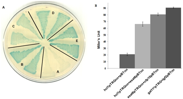
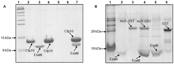
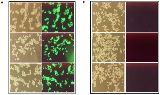
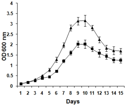
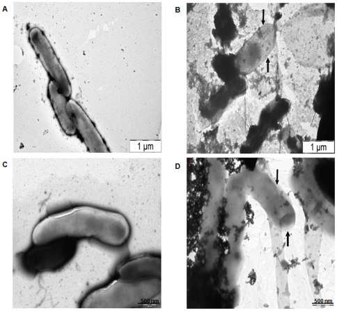
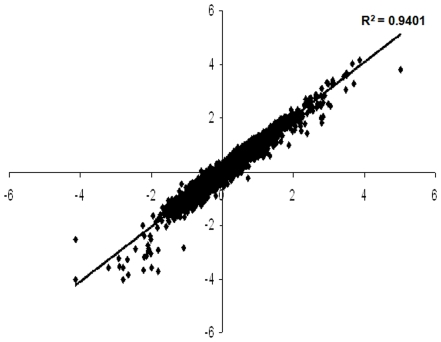
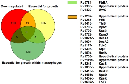
Methodology/principal findings: During our efforts to isolate potential binding partners of key secretory proteins of M. tuberculosis from a human lung protein library, we isolated peptides that strongly bound the virulence determinant protein Esat6. All peptides were less than fifty amino acids in length and the binding was confirmed by in vivo as well as in vitro studies. Curiously, we found all three binders to be unusually rich in phenylalanine, with one of the three peptides a short fragment of the human cytochrome c oxidase-3 (Cox-3). The most accessible of the three binders, named Hcl1, was shown also to bind to the Mycobacterium smegmatis (M. smegmatis) Esat6 homologue. Expression of hcl1 in M. tuberculosis H37Rv led to considerable reduction in growth. Microarray analysis showed that Hcl1 affects a host of key cellular pathways in M. tuberculosis. In a macrophage infection model, the sets expressing hcl1 were shown to clear off M. tuberculosis in much greater numbers than those infected macrophages wherein the M. tuberculosis was not expressing the peptide. Transmission electron microscopy studies of hcl1 expressing M. tuberculosis showed prominent expulsion of cellular material into the matrix, hinting at cell wall damage.

Conclusions/significance: While the debilitating effects of Hcl1 on M. tuberculosis are unrelated and not because of the peptide's binding to Esat6-as the latter is not an essential protein of M. tuberculosis-nonetheless, further studies with this peptide, as well as a closer inspection of the microarray data may shed important light on the suitability of such small phenylalanine-rich peptides as potential drug-like molecules against this pathogen.


Conflict of interest statement

Competing Interests: The authors have declared that no competing interests exist.

Figures

Figure 1
Figure 1. Interaction of Esat6 with human cDNA library (HCL) clones.
(A) BacterioMatch two-hybrid reporter strain was co-transformed with (A) hcl1pTRGnn+pBTnn (negative control); (B) hcl1pTRGnn+esat6pBTnn; (C) hcl2pTRGnn+esat6pBTnn; (D) hcl3pTRGnn+esat6pBTnn; and (E) gal11ppTRG+lgf2pBT (positive control). Two individual colonies from each co-transformant were patched on X-gal indicator plates. (B) Confirmation of the in vivo interaction between Hcl1 and Esat6 using the liquid β-galactosidase assay. Assays were performed in duplicates with appropriate controls. Mean±s.d values are displayed here. A similar pattern was observed each time the experiment was repeated.
Figure 2
Figure 2. In vitro confirmation of the protein-protein interactions.
(A) 16.5% Tricine-SDS polyacrylamide gel stained with coomassie blue showing interaction between purified Esat6 and Cfp10. Lane 1: pre-stained protein marker, Lane 2: purified His-tagged Cfp10; Lane 3: purified FLAG-tagged Esat6; Lane 4: 10 µg Cfp10 alone incubated with Ni-NTA agarose and eluted; Lane 5: 10 µg Esat6 alone incubated with 1% PVP blocked Ni-NTA agarose beads and eluted; Lane 6: empty; Lane 7: 10 µg Cfp10 incubated with Ni-NTA agarose beads and further incubated with 10 µg Esat6, and subsequently eluted. (B) 16.5% Tricine-SDS polyacrylamide gel stained with coomassie blue showing interaction between Esat6 and Hcl1. Lane 1: protein marker; Lane 2: purified His-tagged Esat6; Lane 3: purified Hcl1-GST; Lane 4: 5 µg Esat6 incubated with Ni-NTA agarose beads and further incubated with 5 µg Hcl1-GST, and subsequently eluted; Lane 5: beads initially bound with Esat6 and further incubated with purified GST protein; Lane 6: purified GST protein.
Figure 3
Figure 3. Expression of Hcl1 as a GFP fusion protein in M. tuberculosis.
Three individual colonies each from mycobacteria harbouring either hcl1gfppVV16 (Mtb/GFP-Hcl1) or pVV16 (Mtb/pVV16) were grown till mid-log phase. Cells were examined under a fluorescent microscope. Panel A: Mtb/GFP-Hcl1 cells; Panel B: Mtb/pVV16 cells. First column in each panel shows bright field image of the sample.
Figure 4
Figure 4. Effect of Hcl1 on survival of mycobacteria within macrophages.
THP1 cells were infected with mycobacteria harbouring either hcl1pVV16 (Mtb/Hcl1) or pVV16 (Mtb/pVV16). The figure shows result of CFU counts, immediately after 4 hours of infection (A), called Day 0 and 24 hours post infection (B), called Day 1. Experiment was performed in triplicates and mean±s.d values are displayed here. A significant decline in the survival of mycobacteria carrying hcl1pVV16 was observed in comparison to the vector controls.
Figure 5
Figure 5. Effect of Hcl1 on the growth of M. tuberculosis.
Three individual colonies each from mycobacteria harbouring either hcl1pVV16 (Mtb/Hcl1) or pVV16 (Mtb/pVV16) were picked for growth curve analysis. Optical density of each culture was measured at 600 nm. Mean±s.d values are plotted against time (in Days). A similar growth curve pattern was observed each time the experiment was repeated. Triangular data points represent OD600 values of Mtb/pVV16 samples; Squares represent OD600 values of Mtb/Hcl1 samples.
Figure 6
Figure 6. Effect of Hcl1 on cell shape and morphology of M. tuberculosis.
Transmission electron micrographs of mycobacterial cells harbouring either pVV16 (Mtb/pVV16) or hcl1pVV16 (Mtb/Hcl1). Arrow heads in Panel B and D show the difference in cell morphology of mycobacteria containing Hcl1 when compared with the control (see Panel A and C).
Figure 7
Figure 7. Scatter plot of average Z score values from treated (Mtb/Hcl1) and control (Mtb/pVV16) samples obtained after Z score transformation.
Data points represent the average Z score values of genes obtained from six arrays.
Figure 8
Figure 8. Comparison with Rubin's lists of genes that were found to be upregulated in microarray experiments.
Red circle shows the genes upregulated; yellow and green circles show the genes essential for in vitro growth and for survival of mycobacteria within macrophages, respectively.
Figure 9
Figure 9. Comparison with Rubin's lists of genes that were found to be downregulated in microarray experiments.
Red circle shows the genes upregulated; yellow and green circles show the genes essential for in vitro growth and for survival of mycobacteria within macrophages, respectively.
Figure 10
Figure 10. Relative fold change in the transcript levels of bfrB and groES genes.
Real-time PCR was done using SYBR Green dye. Both genes were found to be significantly downregulated (∼2-fold) in Mtb/Hcl1cells when compared with the mycobacteria harbouring only pVV16. Experiment was performed using two biological replicates and in duplicates.

Similar articles

Cited by

References

    1. Dye C, Scheele S, Dolin P, Pathania V, Raviglione MC. Global burden of tuberculosis: estimated incidence, prevalence, and mortality by country. J Am Med Assoc. 1999;282:677–686. - PubMed
    1. Yew WW, Leung CC. Updates in tuberculosis 2007. Am J Respir Crit Care Med. 2008;177:479–485. - PubMed
    1. Khatri GR. Controlling tuberculosis in India. New Engl J Med. 2002;347:1420–1425. - PubMed
    1. Jain A, Mondal R. Extensively drug-resistant tuberculosis: current challenges and threats. FEMS Immunol Med Microbiol. 2008;53:145–150. - PubMed
    1. Mondal R, Jain A. Extensively Drug-Resistant Mycobacterium tuberculosis, India. Emerg Infect Dis. 2007;13:1429–1431. - PMC - PubMed

Publication types