. 2009 Nov 10;4(11):e7769.
doi: 10.1371/journal.pone.0007769.

Potent inhibition of HIV-1 replication by a Tat mutant

Affiliations

Potent inhibition of HIV-1 replication by a Tat mutant

Luke W Meredith et al. PLoS One. .

Abstract

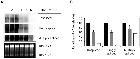
Herein we describe a mutant of the two-exon HIV-1 Tat protein, termed Nullbasic, that potently inhibits multiple steps of the HIV-1 replication cycle. Nullbasic was created by replacing the entire arginine-rich basic domain of wild type Tat with glycine/alanine residues. Like similarly mutated one-exon Tat mutants, Nullbasic exhibited transdominant negative effects on Tat-dependent transactivation. However, unlike previously reported mutants, we discovered that Nullbasic also strongly suppressed the expression of unspliced and singly-spliced viral mRNA, an activity likely caused by redistribution and thus functional inhibition of HIV-1 Rev. Furthermore, HIV-1 virion particles produced by cells expressing Nullbasic had severely reduced infectivity, a defect attributable to a reduced ability of the virions to undergo reverse transcription. Combination of these inhibitory effects on transactivation, Rev-dependent mRNA transport and reverse transcription meant that permissive cells constitutively expressing Nullbasic were highly resistant to a spreading infection by HIV-1. Nullbasic and its activities thus provide potential insights into the development of potent antiviral therapeutics that target multiple stages of HIV-1 infection.


Conflict of interest statement

Competing Interests: The authors have declared that no competing interests exist.

Figures

Figure 1
Figure 1. Nullbasic is a HIV-1 Tat mutant that localizes to the cell cytoplasm.
(A) Amino-acid sequence alignment of Nullbasic (upper rows) against the BH10 clone of Tat (lower rows). Vertical bars indicate amino acid identity and dots indicate engineered substitutions. The solid box highlights the engineered basic domain mutations in Nullbasic, while the dashed box indicates a FLAG epitope tag added to the carboxy terminal. (B) HeLa cells expressing Nullbasic (top row) or Tat-FLAG (botton row) were visualized by confocal microscopy using anti-FLAG/FITC antibodies. Nuclei were stained with DAPI. Images are representative of 6 fields per slide from two independent experiments.
Figure 2
Figure 2. Nullbasic inhibits Tat-mediated transactivation.
Increasing amounts (200 ng, 2 µg and 4 µg) or Nullbasic expression vector were titrated into cells co-transfected with constant amounts of Tat-FLAG plasmid, a luciferase reporter of Tat transactivation (HIV-1 LTR promoter) and a constitutive β-galactosidase expression plasmid (CMV promoter). Luciferase values (CPS, counts per second) were normalized to β-galactosidase activity (µU). Asterisks indicate significant decreases (p<0.05) in transactivation compared to uninhibited Tat-FLAG (first column). Columns represent the means and standard deviations of three independent experiments.
Figure 3
Figure 3. Nullbasic inhibits HIV-1 virion production but not composition.
(A) Virions produced in HEK293T cells co-transfected with 1∶4 molar ratios of HIV-1 plasmid (pGCH) to Tat-FLAG, Nullbasic or empty vector (pcDNA3.1+; “No Tat”) plasmids were assayed for capsid (CA) and reverse transcriptase (RT) concentrations. The CA (black columns) and RT (white columns) concentrations are shown. Columns represent the means and standard deviations of four independent experiments and are expressed as a percentage of the “No Tat” sample. Asterisks indicate significant differences (p<0.025, Welch's t-test) in CA and RT concentrations between Nullbasic and Tat-FLAG samples. (B) HEK293T cells transfected with a reporter plasmid that expresses Rev-independent Env from the CMV/LTR hybrid promoter of pGCH were co-expressed with (lane 2) or without (lane 1) Nullbasic. Env and Nullbasic were detected by immunoblotting with anti-gp120 and anti-FLAG antibodies, respectively. (C) Cell lysates from HEK293T cells expressing pGCH provirus alone (lane 1) or co-expressing either Tat-FLAG (lanes 2 and 3; 1∶2 and 1∶4 molar ratios, respectively) or Nullbasic (lanes 4 and 5; 1∶2 and 1∶4 molar ratios, respectively) were immunoblotted with a monoclonal antibody against HIV-1 SU (upper panel), anti-serum against HIV (middle panel) and a monoclonal antibody against FLAG (lower panel). A β-galactosidase expression plasmid was included in all transfections and western blotting was performed on lysates normalised for β-galactosidase activity. (D) Culture supernatants were collected from HEK293T cells expressing pGCH alone (lane 1) or co-expressing either Nullbasic (lane 2) or Tat-FLAG (lane 3). Virions in the supernatants were concentrated by ultracentrifugation before samples containing 50 ng of total CA were immunoblotted with anti-gp120 and anti-CA antibodies. Data in B, C and D are representative of three independent experiments. (E) Packaged genomic RNA was isolated from virions produced by HEK293T cells expressing pGCH co-transfected with empty vector (pcDNA3.1+; “No Tat”) or co-expressing Tat-FLAG or Nullbasic before being quantitated by RT-PCR. The means and standard deviations of two experiments performed in duplicate on independent virus stocks are shown, with values expressed as a percentage of the “No Tat” sample.
Figure 4
Figure 4. Nullbasic downregulates the levels of unspliced and singly-spliced HIV-1 mRNA expressed in cells.
(A) Northern blot analysis of total RNA from HEK293T cells expressing pGCH provirus co-transfected with empty vector (pcDNA3.1+; “No Tat”, lane 1) or co-expressing increasing amounts of Tat-FLAG (lanes 2 and 3; 1∶2 and 1∶4 molar ratios, respectively) or Nullbasic (lanes 4 and 5; 1∶2 and 1∶4 molar ratios, respectively). An untransfected control was also included (lane 6). Unspliced, singly-spliced and multiply-spliced HIV-1 mRNA were detected with a single HIV-1-specific probe. 28S and 18S ribosomal RNA (rRNA) species demonstrate equal sample loading. Data are representative of four independent experiments. (B) Total RNA was extracted from HEK293T cells expressing pGCH co-transfected with empty vector (black bars) or co-expressing Tat-FLAG (gray bars) or Nullbasic (white bars) before RT-PCR reactions were performed using primers specific to unspliced, singly-spliced and multiply-spliced viral mRNA. Tat-FLAG and Nullbasic plasmids were transfected at 2∶1 molar ratios with respect to pGCH. The means and standard deviations of duplicate assays in three independent experiments are shown, with values for each mRNA class expressed as a percentage of the pGCH alone sample.
Figure 5
Figure 5. Nullbasic alters the subcellular localization of HIV-1 Rev.
HeLa cells expressing a Myc-Rev fusion protein alone (top row), Myc-Rev with Nullbasic (middle row) or Myc-Rev with Tat-FLAG (bottom row) were visualized by confocal microscopy using anti-Myc/Cy3 and anti-FLAG/FITC antibodies. Nuclei were stained with DAPI. The total amounts of transfected plasmids in each experiment were normalized with empty vector (pcDNA3.1+). Images are representative of at least five fields per slide from three independent experiments.
Figure 6
Figure 6. Nullbasic inhibits the RNA export function of provirus-expressed Rev.
(A) Schematic diagram of the Rev-dependent CAT expression cassette in plasmid pDM128. The chloramphenicol acetyltransferase (CAT) gene exists within an artificial intron bounded by splice donor (SD) and splice acceptor (SA) sequences. Successful expression of CAT protein requires binding of Rev to the Rev response element (RRE) to avoid intron splicing and to enable nuclear export of the CAT mRNA transcript. SV40: simian virus 40; 3′ LTR: HIV-1 long terminal repeat (contains polyadenylation signal). (B) HEK293T cells were co-transfected with pDM128 and pGCH provirus alone (column 2), Myc-Rev plasmid alone (column 4) or along with Nullbasic plasmid (columns 3 and 5). Empty vector (pcDNA3.1+) was used to normalize the total amount of transfected plasmids. A pDM128 only transfection was included as a negative control (column 1) and a constitutive β-galactosidase plasmid was included in all transfections to account for variations in transfection efficiencies. CAT expression was measured by ELISA and is expressed relative to β-galactosidase activity (ng/mU). Columns represent means and standard deviations of three independent experiments. (C) Lysates from cells expressing a Rev-independent Gag-FLAG fusion protein co-transfected with empty vector (lane 1) or co-expressing either Tat-FLAG (lanes 2 and 3; 1∶2 and 1∶4 molar ratio, respectively) or Nullbasic (lanes 4 and 5; 1∶2 and 1∶4 molar ratio, respectively) were immunoblotted with anti-FLAG antibody to detect all three proteins. The white band centers in lanes 3 and 5 are due to luminol precipitation, indicative of very high amounts of Tat-FLAG and Nullbasic protein, respectively, on the membrane. Data are representative of three independent experiments.
Figure 7
Figure 7. Nullbasic inhibits HIV-1 infectivity and endogenous reverse transcription.
(A) The infectivity of HIV-1 produced by HEK293T cells expressing pGCH provirus co-transfected with empty vector (pcDNA3.1+) or co-expressing increasing amounts of Nullbasic or Tat-FLAG (at 1∶2 and 1∶4 molar ratios) were determined by the MAGI assay. The indicator cells were infected with virion samples equalized for reverse transcriptase activity before cell lysates were assayed for β-galactosidase production 48 h later. Columns represent the means and standard deviations of three independent experiments and are expressed as a percentage of the “No Tat” sample (column 1). Asterisks indicate significant differences (p<0.05) in infectivities compared to the “No Tat” sample. (B) Detergent-free endogenous reverse transcription assays were performed with the same virus samples as in A. The amount of negative-strand strong-stop DNA (–ssDNA) was quantitated by PCR, and normalized to the RT activity in each sample. The asterisk indicates a significant difference (p<0.05) in –ssDNA synthesis relative to the “No Tat” sample, and columns represent the means and standard deviations of duplicate assays in three independent experiments.
Figure 8
Figure 8. Expression of Nullbasic-EGFP in permissive target cells suppresses HIV-1 infection and spread.
(A) The ability of Nullbasic-EGFP to inhibit HIV-1 infectivity was compared to Nullbasic in a MAGI assay similar to Figure 7A (using a 1∶2 molar ratio only). Columns represent the means and standard deviations of three independent experiments and are expressed as a percentage of the “No Tat” (pcDNA3.1+ co-transfected) sample. (B) Cell surface expression of transgenic CD4 receptors was quantitated in MAGI (black) and MAGI/Nullbasic-EGFP (red) cells by flow cytometry using an anti-CD4 monoclonal antibody. (C) Cell surface expression of endogenous CXCR4 receptors was quantitated in MAGI (black) and MAGI/Nullbasic-EGFP (red) cells by flow cytometry using an anti-CXCR4 monoclonal antibody. (D) The MAGI (black circles), MAGI/EGFP (black diamonds) and MAGI/Nullbasic-EGFP (white diamonds) cell lines were infected with equal amounts of pGCH-derived virions (500 ng CA-equivalent per 106 cells) before viral replication was monitored over a 14-day period. Log values of virion CA levels measured in the culture supernatants are shown plotted against time. Obelisks (†) indicate observations of syncytia and cell death in the MAGI and MAGI/EGFP cell lines at 9 days post-infection. Data points represent the means and standard deviations of triplicate assays in two independent experiments.

Similar articles

Cited by

References

    1. Dayton AI, Sodroski JG, Rosen CA, Goh WC, Haseltine WA. The trans-activator gene of the human T cell lymphotropic virus type III is required for replication. Cell. 1986;44:941–947. - PubMed
    1. Hauber J, Perkins A, Heimer EP, Cullen BR. Trans-activation of human immunodeficiency virus gene expression is mediated by nuclear events. Proc Natl Acad Sci U S A. 1987;84:6364–6368. - PMC - PubMed
    1. Hauber J, Malim MH, Cullen BR. Mutational analysis of the conserved basic domain of human immunodeficiency virus tat protein. J Virol. 1989;63:1181–1187. - PMC - PubMed
    1. Ruben S, Perkins A, Purcell R, Joung K, Sia R, et al. Structural and functional characterization of human immunodeficiency virus tat protein. J Virol. 1989;63:1–8. - PMC - PubMed
    1. Berkhout B, Silverman RH, Jeang KT. Tat trans-activates the human immunodeficiency virus through a nascent RNA target. Cell. 1989;59:273–282. - PubMed

Publication types