

Identification of a dual-specific T cell epitope of the hemagglutinin antigen of an h5 avian influenza virus in chickens
- PMID: 19901990
- PMCID: PMC2770124
- DOI: 10.1371/journal.pone.0007772
Identification of a dual-specific T cell epitope of the hemagglutinin antigen of an h5 avian influenza virus in chickens
Abstract
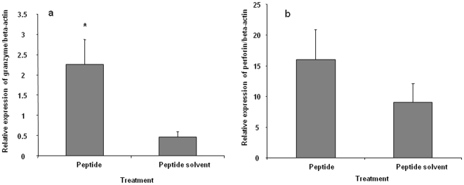
Avian influenza viruses (AIV) of the H5N1 subtype have caused morbidity and mortality in humans. Although some migratory birds constitute the natural reservoir for this virus, chickens may play a role in transmission of the virus to humans. Despite the importance of avian species in transmission of AIV H5N1 to humans, very little is known about host immune system interactions with this virus in these species. The objective of the present study was to identify putative T cell epitopes of the hemagglutinin (HA) antigen of an H5 AIV in chickens. Using an overlapping peptide library covering the HA protein, we identified a 15-mer peptide, H5(246-260,) within the HA1 domain which induced activation of T cells in chickens immunized against the HA antigen of an H5 virus. Furthermore, H5(246-260) epitope was found to be presented by both major histocompatibility complex (MHC) class I and II molecules, leading to activation of CD4+ and CD8+ T cell subsets, marked by proliferation and expression of interferon (IFN)-gamma by both of these cell subsets as well as the expression of granzyme A by CD8+ T cells. This is the first report of a T cell epitope of AIV recognized by chicken T cells. Furthermore, this study extends the previous finding of the existence of dual-specific epitopes in other species to chickens. Taken together, these results elucidate some of the mechanisms of immune response to AIV in chickens and provide a platform for creation of rational vaccines against AIV in this species.
Conflict of interest statement
Figures

Similar articles
-
Identification of novel avian influenza virus derived CD8+ T-cell epitopes.PLoS One. 2012;7(2):e31953. doi: 10.1371/journal.pone.0031953. Epub 2012 Feb 23. PLoS One. 2012. PMID: 22384112 Free PMC article.
-
Improved immune responses against avian influenza virus following oral vaccination of chickens with HA DNA vaccine using attenuated Salmonella typhimurium as carrier.Comp Immunol Microbiol Infect Dis. 2012 Sep;35(5):417-27. doi: 10.1016/j.cimid.2012.03.007. Epub 2012 Apr 16. Comp Immunol Microbiol Infect Dis. 2012. PMID: 22512819
-
Construction of a recombinant duck enteritis virus (DEV) expressing hemagglutinin of H5N1 avian influenza virus based on an infectious clone of DEV vaccine strain and evaluation of its efficacy in ducks and chickens.Virol J. 2015 Aug 13;12:126. doi: 10.1186/s12985-015-0354-9. Virol J. 2015. PMID: 26263920 Free PMC article.
-
Prime-Boost Vaccination With a Novel Hemagglutinin Protein Produced in Bacteria Induces Neutralizing Antibody Responses Against H5-Subtype Influenza Viruses in Commercial Chickens.Front Immunol. 2019 Sep 4;10:2006. doi: 10.3389/fimmu.2019.02006. eCollection 2019. Front Immunol. 2019. PMID: 31552018 Free PMC article.
-
Development and use of fowlpox vectored vaccines for avian influenza.Ann N Y Acad Sci. 2006 Oct;1081:193-201. doi: 10.1196/annals.1373.023. Ann N Y Acad Sci. 2006. PMID: 17135511 Review.
Cited by
-
Progress on chicken T cell immunity to viruses.Cell Mol Life Sci. 2019 Jul;76(14):2779-2788. doi: 10.1007/s00018-019-03117-1. Epub 2019 May 17. Cell Mol Life Sci. 2019. PMID: 31101935 Free PMC article. Review.
-
Identification of a Highly Conserved Epitope on Avian Influenza Virus Non-Structural Protein 1 Using a Peptide Microarray.PLoS One. 2016 Mar 3;11(3):e0149868. doi: 10.1371/journal.pone.0149868. eCollection 2016. PLoS One. 2016. PMID: 26938453 Free PMC article.
-
An infected chicken kidney cell co-culture ELISpot for enhanced detection of T cell responses to avian influenza and vaccination.J Immunol Methods. 2015 Jan;416:40-8. doi: 10.1016/j.jim.2014.10.012. Epub 2014 Nov 7. J Immunol Methods. 2015. PMID: 25450002 Free PMC article.
-
Evolutionary pressures rendered by animal husbandry practices for avian influenza viruses to adapt to humans.iScience. 2022 Mar 1;25(4):104005. doi: 10.1016/j.isci.2022.104005. eCollection 2022 Apr 15. iScience. 2022. PMID: 35313691 Free PMC article. Review.
-
Structures of the MHC-I molecule BF2*1501 disclose the preferred presentation of an H5N1 virus-derived epitope.J Biol Chem. 2020 Apr 17;295(16):5292-5306. doi: 10.1074/jbc.RA120.012713. Epub 2020 Mar 9. J Biol Chem. 2020. PMID: 32152225 Free PMC article.
References
Publication types
MeSH terms
Substances
LinkOut - more resources
Full Text Sources
Other Literature Sources
Medical
Research Materials
Miscellaneous
