

Prognostic value of microvascular density in dukes a and B (t1-t4, n0, m0) colorectal carcinomas
- PMID: 19902004
- PMCID: PMC2774471
- DOI: 10.1155/2009/679830
Prognostic value of microvascular density in dukes a and B (t1-t4, n0, m0) colorectal carcinomas
Abstract
Background: Aproximatelly 30% of patients operated on for colorectal cancer (CRC), with an expectedly favourable prognosis (Dukes A-B/T1-T4, N0, M0) suffer recurrence and/or die.
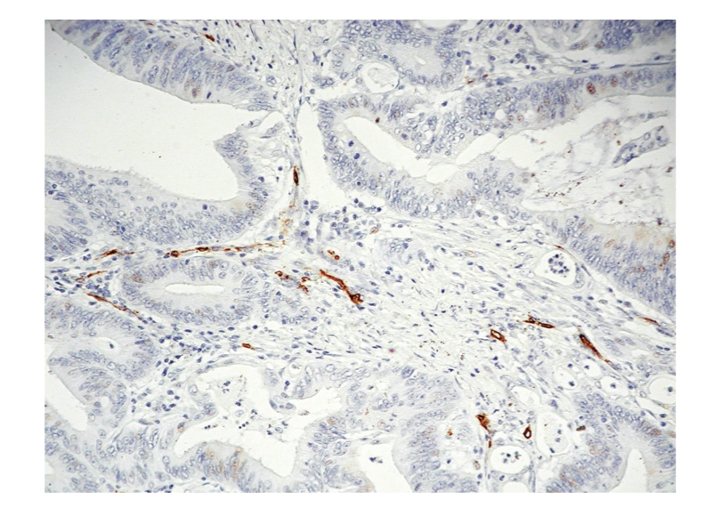
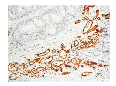
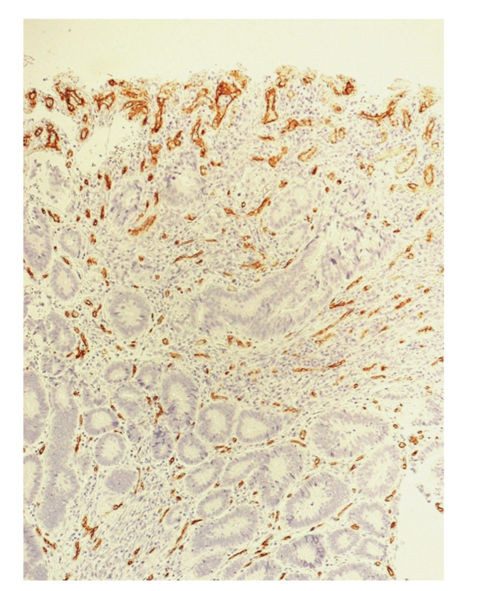
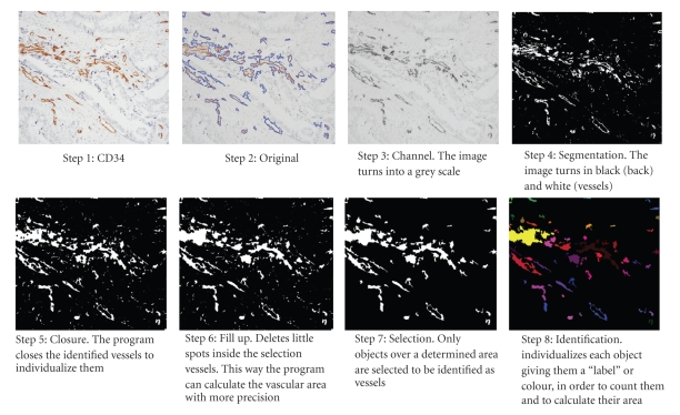
Method: In order to determine if tumor microvascular density (MVD) is a prognostic factor in CRC, samples from tumors of 104 Dukes A-B CRC patients were retrospectively studied. Immunohistochemistry was performed for anti-CD34 antibody to visualize tumor vascularisation. MVD was expressed as the total number of vessels and as the percentage of microvascular area. We calculated MVD with a morphometry program and performed descriptive, bivariate, and survival statistics.
Results: The mean number of vessels was 37.37/200x field, and the mean vascular area was the 3.972%. 30% of the patients with < 37 vessels/field, and 21% of the patients with > 37 vessels/field, experienced recurrence/death. The 35% of patients with < 4% of vascular area died following recurrence, compared with 14% of patients with > or =4% of vascular area. These differences in % of vascular area were statistically significant.
Conclusion: MVD expressed as the total number of vessels had no a statistically significant influence on the evolution of CRC. However, neoplasias with a greater % of vascular were associated to a better outcome.
Figures
Similar articles
-
Prognostic value of lymph node micrometastasis in patients with colorectal cancer in Dukes stages A and B (T1-T4, N0, M0).Rev Esp Enferm Dig. 2010 Mar;102(3):176-86. doi: 10.4321/s1130-01082010000300004. Rev Esp Enferm Dig. 2010. PMID: 20373832 English, Spanish.
-
Immunohistochemical expression of vascular endothelial growth factor and microvessel counting as prognostic indicators in node-negative colorectal cancer.Tumour Biol. 2005 Jan-Feb;26(1):1-8. doi: 10.1159/000084180. Epub 2005 Feb 28. Tumour Biol. 2005. PMID: 15741766
-
The CD34-microvascular density in colorectal cancer patients.J BUON. 2012 Jan-Mar;17(1):97-101. J BUON. 2012. PMID: 22517700
-
Prognostic value of MVD, LVD and vascular invasion in lymph node-negative colon cancer.Hepatogastroenterology. 2013 May;60(123):432-8. doi: 10.5754/hge12826. Hepatogastroenterology. 2013. PMID: 23321007
-
Both high intratumoral microvessel density determined using CD105 antibody and elevated plasma levels of CD105 in colorectal cancer patients correlate with poor prognosis.Br J Cancer. 2003 May 6;88(9):1424-31. doi: 10.1038/sj.bjc.6600874. Br J Cancer. 2003. PMID: 12778073 Free PMC article.
Cited by
-
Increased chemoresistance via Snail-Raf kinase inhibitor protein signaling in colorectal cancer in response to a nicotine derivative.Oncotarget. 2016 Apr 26;7(17):23512-20. doi: 10.18632/oncotarget.8049. Oncotarget. 2016. PMID: 26992205 Free PMC article.
-
Role of VEGF, CD105, and CD31 in the Prognosis of Colorectal Cancer Cases.J Gastrointest Cancer. 2019 Mar;50(1):23-34. doi: 10.1007/s12029-017-0014-y. J Gastrointest Cancer. 2019. PMID: 29110224
-
Polymorphisms in XPD and ERCC1 Associated with Colorectal Cancer Outcome.Int J Mol Sci. 2013 Feb 19;14(2):4121-34. doi: 10.3390/ijms14024121. Int J Mol Sci. 2013. PMID: 23429196 Free PMC article.
-
Quantification of tumour budding, lymphatic vessel density and invasion through image analysis in colorectal cancer.J Transl Med. 2014 Jun 1;12:156. doi: 10.1186/1479-5876-12-156. J Transl Med. 2014. PMID: 24885583 Free PMC article.
-
Recurrences in stage II rectal carcinoma after curative resection alone: from the viewpoint of angiogenesis.World J Surg Oncol. 2016 Apr 22;14:122. doi: 10.1186/s12957-016-0877-6. World J Surg Oncol. 2016. PMID: 27102733 Free PMC article.
References
-
- Fielding P. Staging systems. In: Cohen A, Vimawer S, editors. Cancer of the Colon, Rectum, and Anus. New York, NY, USA: McGraw-Hill; 1995. p. 207.
-
- Itzkowitz SH. Cancer gastrointestinal: cancer de colon y recto. In: Wilcox CM, editor. Digestive Diseases Self Education Program. Barcelona, Spain: Medical Trends; 2001. p. 25.
-
- Hermanek P. pTNM and residual tumor classifications: problems of assesment and prognostic significance. The World Journal of Surgery. 1995;19:180–190. - PubMed
-
- Bilchik AJ, Saha S, Wiese D, et al. Molecular staging of early colon cancer on the basis of sentinel node analysis: a multicenter phase II trial. Journal of Clinical Oncology. 2001;19(4):1128–1136. - PubMed
-
- Ovaska J, Järvinen H, Kujari H, et al. Follow-up of patients operated on for colorectal carcinoma. American Journal of Surgery. 1990;159(6):593–596. - PubMed
LinkOut - more resources
Full Text Sources
