. 2010 Feb;67(3):433-43.
doi: 10.1007/s00018-009-0190-4. Epub 2009 Nov 10.

Visualization of subcellular NAD pools and intra-organellar protein localization by poly-ADP-ribose formation

Affiliations

Visualization of subcellular NAD pools and intra-organellar protein localization by poly-ADP-ribose formation

Christian Dölle et al. Cell Mol Life Sci. 2010 Feb.

Abstract

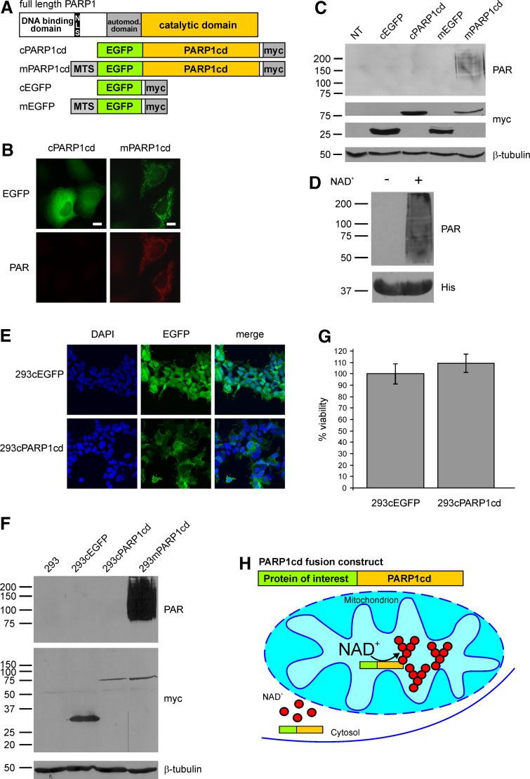
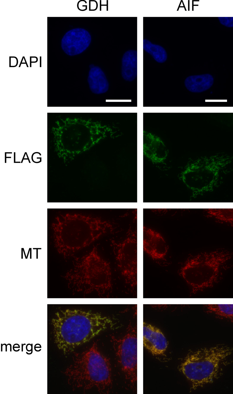
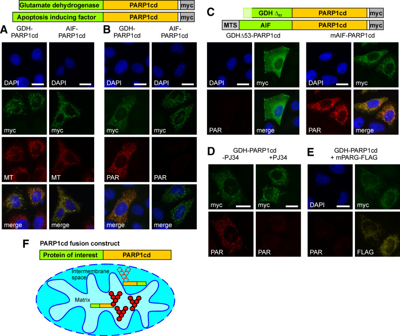
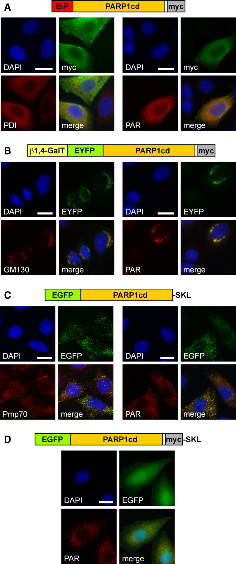
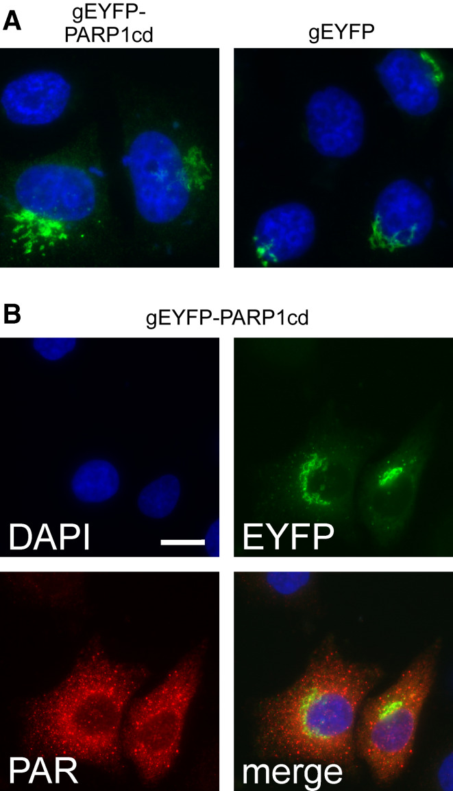
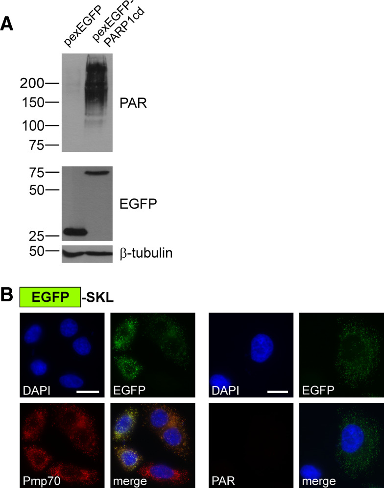
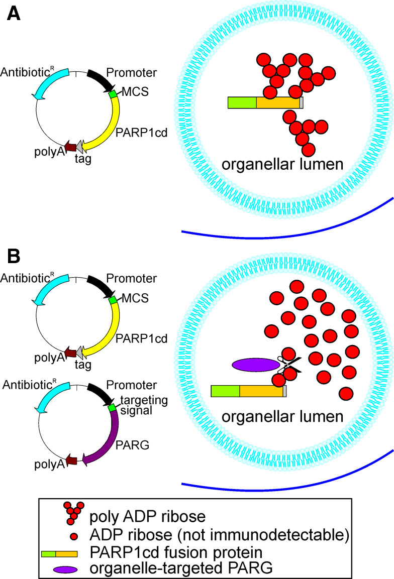
Poly-ADP-ribose polymerases (PARPs) use NAD(+) as substrate to generate polymers of ADP-ribose. We targeted the catalytic domain of human PARP1 as molecular NAD(+) detector into cellular organelles. Immunochemical detection of polymers demonstrated distinct subcellular NAD(+) pools in mitochondria, peroxisomes and, surprisingly, in the endoplasmic reticulum and the Golgi complex. Polymers did not accumulate within the mitochondrial intermembrane space or the cytosol. We demonstrate the suitability of this compartment-specific NAD(+) and poly-ADP-ribose turnover to establish intra-organellar protein localization. For overexpressed proteins, genetically endowed with PARP activity, detection of polymers indicates segregation from the cytosol and consequently intra-organellar residence. In mitochondria, polymer build-up reveals matrix localization of the PARP fusion protein. Compared to presently used fusion tags for subcellular protein localization, these are substantial improvements in resolution. We thus established a novel molecular tool applicable for studies of subcellular NAD metabolism and protein localization.


Figures

Fig. 1
Fig. 1
Mitochondrial, but not cytosolic overexpression of PARP1cd results in immunodetectable PAR accumulation. a Molecular architecture of poly-ADP-ribose polymerase 1 and the generated PARP1cd and EGFP fusion constructs. MTS Mitochondrial targeting sequence, NLS nuclear localization signal. b Fluorescence micrographs of HeLa S3 cells subjected to PAR immunocytochemistry 24 h after transient transfection with vectors encoding cytosolic or mitochondrial EGFP-PARP1cd (cPARP1cd and mPARP1cd). Protein expression was monitored by the intrinsic fluorescence of the EGFP portion of the constructs. Bar 10 μm. c PAR immunoblot analysis of lysates from HeLa S3 cells expressing cPARP1cd or mPARP1cd or the respective constructs lacking the PARP1cd portion (cEGFP and mEGFP). Overexpression of the proteins was detected by the C-terminal myc-tag. As observed in (b), PAR formation was only detected in cells expressing mPARP1cd. Loading control: β-tubulin. d Addition of NAD+ (1 mM) to bacterially expressed human PARP1cd (amino acids 652–1014) led to automodification of the protein as visualized by PAR immunoblot analysis. The bottom part shows the immunodetection of the protein’s 6xHis-tag. e Stably transfected 293 cells expressing cytosolic EGFP (293cEGFP) or cytosolic EGFP-PARP1cd (293cPARP1cd). Protein expression as monitored by the intrinsic fluorescence of the EGFP portion of the constructs is detectable in all cells. f Immunoblot analyses of lysates from parental and stably transfected 293 cell lines. Expression of cEGFP, cPARP1cd and mPARP1cd was detected by the C-terminal myc epitope. Mitochondrial and cytosolic PARP1cd fusion proteins were expressed at similar levels. PAR immunoblot analysis using 10H antibody revealed detectable PAR formation only in cells expressing mitochondrial PARP1cd. Loading control: β-tubulin. g Constitutive expression of cPARP1cd does not affect cell viability. Cells were seeded into 96-well plates and the viability was determined by MTT assay. Viability of 293cPARP1cd was related to control 293cEGFP cells set to 100%. Data are shown as mean ± SE of four experiments, each performed in triplicate. h Schematic representation of the observed differences in PAR accumulation mediated by targeted overexpression of PARP1cd fusion proteins. Cytosolic PARP1cd does not give rise to immunodetectable PAR. NAD and ADP-ribose are not recognized by the PAR-specific antibodies. The presence of PARP1cd within mitochondria leads to extensive PAR formation
Fig. 2
Fig. 2
Association of GDH- and AIF-FLAG constructs with mitochondria. HeLa S3 cells were transiently transfected with vectors encoding GDH and AIF as FLAG tagged proteins. After 24 h cells were stained with MitoTracker (MT) and subjected to FLAG-immunocytochemistry. The fluorescence micrographs show nuclei (DAPI), expressed recombinant proteins (FLAG), and mitochondria (MT). Bar 10 μm
Fig. 3
Fig. 3
Submitochondrial localization of PARP1cd fusion proteins based on PAR accumulation in the matrix, but not the intermembrane space. a GDH-PARP1cd and AIF-PARP1cd fusion proteins localize to mitochondria. HeLa S3 cells were transiently transfected with vectors encoding the indicated mitochondrial PARP1cd fusion proteins. After 24 h, cells were stained with MitoTracker (MT) and subjected to myc-immunocytochemistry. The fluorescence micrographs show nuclei (DAPI), expressed recombinant proteins (myc), and mitochondria (MT). Bar 10 μm. b Robust PAR accumulation is restricted to the mitochondrial matrix. After transient transfection of vectors encoding GDH-PARP1cd and AIF-PARP1cd, HeLa S3 cells were subjected to immunocytochemistry using myc- and PAR-specific antibodies. The fluorescence micrographs show nuclei (DAPI), expressed recombinant proteins (myc), and generated PAR. Bar 10 μm. c Artificial re-targeting of mitochondrial PARP1cd fusion proteins confirms matrix localization to be essential for PAR accumulation. HeLa S3 cells were transfected with vectors encoding the indicated PARP1cd fusion constructs (matrix-targeted mAIF-PARP1cd and GDHΔ53-PARP1cd lacking the mitochondrial targeting sequence) and subjected to immunocytochemistry as in (b). Bar 10 μm. d PAR accumulation requires PARP1cd catalytic activity. HeLa S3 cells transiently transfected with a GDH-PARP1cd encoding plasmid were cultured in presence (+) or absence (−) of the PARP1 inhibitor PJ34 (5 μM). Cells were subjected to PAR- and myc-immunocytochemistry 24 h post transfection as in (b). Bar 10 μm. e Co-expression of matrix-targeted PARG (mPARG) eliminates GDH-PARP1cd-mediated PAR accumulation. HeLa S3 cells were co-transfected with vectors encoding GDH-PARP1cd and a FLAG-tagged mitochondrial PARG construct (mPARG). The fluorescence micrographs show nuclei (DAPI), GDH-PARP1cd (myc), mPARG (FLAG), and polymers (PAR). Bar 10 μm. f Schematic representation of the principle to distinguish between mitochondrial matrix and intermembrane space protein localization. If PARP1cd fusion proteins are directed into the matrix, PAR accumulation (aggregates of red spheres) is readily observed, whereas PAR is virtually not detectable (dotted spheres) for fusion proteins residing in the intermembrane space
Fig. 4
Fig. 4
Targeted expression of PARP1cd to the lumen of the endoplasmic reticulum, the Golgi apparatus or peroxisomes results in detectable PAR formation within these organelles. a HeLa S3 cells transfected with a vector encoding the first 100 amino acids of binding immunoglobulin protein (BiP) N-terminally fused to PARP1cd (BiP-PARP1cd) were subjected to immunocytochemistry after 24 h. The overexpressed protein (myc) co-localized with the ER marker protein disulfide isomerase, PDI (left), and mediated PAR accumulation (right). Bar 10 μm. b HeLa S3 cells were transfected with a vector encoding a Golgi-targeted EYFP-PARP1cd fusion construct, gEYFP-PARP1cd. The intrinsic EYFP-fluorescence of the overexpressed recombinant protein localized to the Golgi complex as revealed by immunostaining of the Golgi-specific marker protein GM130 (left). PAR accumulation (right) was observed in cells overexpressing gEYFP-PARP1cd. Bar 10 μm. c HeLa S3 cells were transiently transfected with a vector encoding EGFP-PARP1cd harboring the C-terminal tripeptide SKL for peroxisomal targeting. Cells were subjected to immunocytochemistry after 24 h using antibodies against the peroxisomal marker protein Pmp70 (left). The overexpressed protein led to PAR accumulation within peroxisomes (right). Expression of the recombinant protein was monitored by its intrinsic EGFP fluorescence. Bar 10 μm. d An additional myc epitope, immediately preceding the C-terminal SKL signal, perturbed peroxisomal localization and led to predominant cytoplasmic distribution of the overexpressed protein. However, accumulation of PAR in peroxisomes revealed that the protein was still partially localized within the organelles
Fig. 5
Fig. 5
Golgi-targeted PARP1cd fusion proteins give rise to PAR-positive cytoplasmic vesicles. a The Golgi-targeted EYFP-PARP1cd fusion protein exhibits the same subcellular distribution as an established Golgi-EYFP construct carrying the same targeting sequence. The proteins were detected by their intrinsic fluorescence, nuclei were stained with DAPI. b HeLa S3 cells expressing a Golgi-targeted PARP1cd fusion construct displayed consistent PAR immunoreactivity as revealed by immunocytochemistry. While the protein was detected in Golgi structures by its intrinsic EYFP fluorescence, the PAR signal most often did not co-localize with the EYFP signal, but was detected in cytoplasmic vesicles. Note that in part of the transfected cells both the protein and the PAR co-localized within the Golgi complex (cf. Fig. 3b). Bar 10 μm
Fig. 6
Fig. 6
Detection of peroxisomal protein localization. a PAR immunoblot analysis of lysates from HeLa S3 cells transiently expressing EGFP or EGFP-PARP1cd targeted to peroxisomes as indicated. PAR accumulation was detected only in lysates from cells expressing the pexEGFP-PARP1cd protein. Each lane was loaded with 70 μg of total protein. The overexpressed proteins were detected using an antibody recognizing the EGFP portion, β-tubulin served as loading control. b HeLa S3 cells transiently expressing EGFP targeted to the peroxisomes by a C-terminal SKL sequence were subjected to immunocytochemistry to detect the endogenous marker Pmp70 (left) or PAR (right). As monitored by its intrinsic EGFP fluorescence the protein co-localized with peroxisomal structures (Pmp70). However, the cells were negative for PAR immunoreactivity. Bar 10 μm
Fig. 7
Fig. 7
Schematic overview of Poly-ADP-Ribose Assisted Protein Localization AssaY (PARAPLAY). a To generate a construct encompassing the protein of interest in fusion with PARP1cd an expression vector (left) can be used harboring a multiple cloning site (MCS) to insert the cDNA encoding the protein of interest upstream of the PARP1cd cDNA. An optional C-terminal tag for protein detection may be included. Transfection of cells with the resultant vector leads to robust PAR accumulation, if the overexpressed protein is targeted to an organellar lumen (right). b Co-transfection of cells with the PARP1cd fusion construct and an organelle-targeted PARG may be conducted to confirm the results. PARG cleaves PAR to ADP ribose monomers, which are not detectable by PAR immunochemistry. Therefore, if the PARP1cd fusion protein resides within the same organelle as the targeted PARG, PAR accumulation is not detectable any more

Similar articles

Cited by

References

    1. Simpson JC, Pepperkok R. The subcellular localization of the mammalian proteome comes a fraction closer. Genome Biol. 2006;7:222. doi: 10.1186/gb-2006-7-6-213. - DOI - PMC - PubMed
    1. Huh WK, Falvo JV, Gerke LC, Carroll AS, Howson RW, Weissman JS, O’Shea EK. Global analysis of protein localization in budding yeast. Nature. 2003;425:686–691. doi: 10.1038/nature02026. - DOI - PubMed
    1. Foster LJ, de Hoog CL, Zhang Y, Zhang Y, Xie X, Mootha VK, Mann M. A mammalian organelle map by protein correlation profiling. Cell. 2006;125:187–199. doi: 10.1016/j.cell.2006.03.022. - DOI - PubMed
    1. O’Rourke NA, Meyer T, Chandy G. Protein localization studies in the age of ‘Omics’. Curr Opin Chem Biol. 2005;9:82–87. doi: 10.1016/j.cbpa.2004.12.002. - DOI - PubMed
    1. Neupert W, Herrmann JM. Translocation of proteins into mitochondria. Annu Rev Biochem. 2007;76:723–749. doi: 10.1146/annurev.biochem.76.052705.163409. - DOI - PubMed

MeSH terms

LinkOut - more resources