. 2010 Feb;53(2):378-88.
doi: 10.1007/s00125-009-1529-y. Epub 2009 Nov 10.

Impaired wound healing in mouse models of diabetes is mediated by TNF-alpha dysregulation and associated with enhanced activation of forkhead box O1 (FOXO1)

Affiliations

Impaired wound healing in mouse models of diabetes is mediated by TNF-alpha dysregulation and associated with enhanced activation of forkhead box O1 (FOXO1)

M F Siqueira et al. Diabetologia. 2010 Feb.

Abstract

Aims/hypothesis: The role of TNF-alpha in impaired wound healing in diabetes was examined by focusing on fibroblasts.

Methods: Small excisional wounds were created in the db/db mice model of type 2 diabetes and normoglycaemic littermates, and in a streptozotocin-induced type 1 diabetes mouse model and control mice. Fibroblast apoptosis was measured by the TUNEL assay, proliferation by detection of proliferating cell nuclear antigen, and forkhead box O1 (FOXO1) activity by DNA binding and nuclear translocation. TNF-alpha was specifically inhibited by pegsunercept.

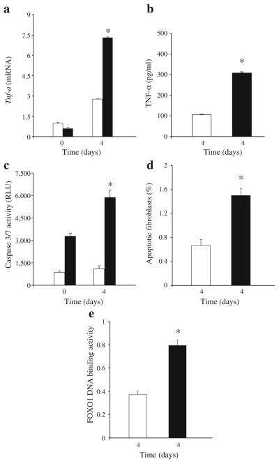
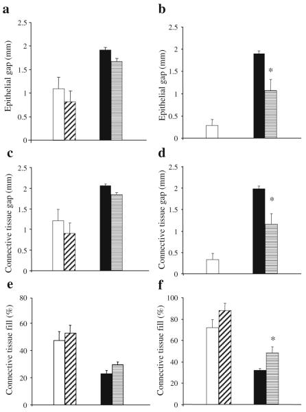
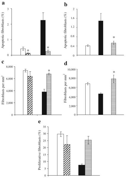
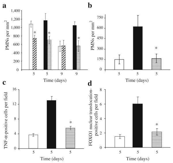
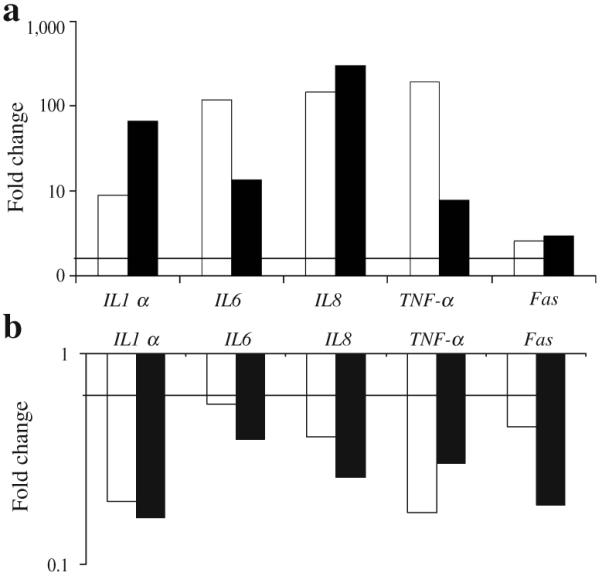
Results: Diabetic wounds had increased TNF-alpha, fibroblast apoptosis, caspase-3/7 activity and activation of the pro-apoptotic transcription factor FOXO1, and decreased proliferating cell nuclear antigen positive fibroblasts (p < 0.05). TNF-alpha inhibition improved healing in the diabetic mice and increased fibroblast density. This may be explained by a decrease in fibroblast apoptosis and increased proliferation when TNF-alpha was blocked (p < 0.05). Although decreased fibroblast proliferation and enhanced FOXO1 activity were investigated in type 2 diabetes, they may also be implicated in type 1 diabetes. In vitro, TNF-alpha enhanced mRNA levels of gene sets related to apoptosis and Akt and p53 but not mitochondrial or cell-cycle pathways. FOXO1 small interfering RNA reduced gene sets that regulate apoptosis, Akt, mitochondrial and cell-cycle pathways. TNF-alpha also increased genes involved in inflammation, cytokine, Toll-like receptor and nuclear factor-kB pathways, which were significantly reduced by FOXO1 knockdown.

Conclusions/interpretation: These studies indicate that TNF-alpha dysregulation in diabetic wounds impairs healing, which may involve enhanced fibroblast apoptosis and decreased proliferation. In vitro, TNF-alpha induced gene sets through FOXO1 that regulate a number of pathways that could influence inflammation and apoptosis.


Figures

Fig. 1
Fig. 1
Diabetes increases TNF-α mRNA and protein levels, fibroblast apoptosis, caspase-3/7 activity and FOXO1 DNA binding in healing wounds. A 1.5 mm excisional full thickness wound was created in the scalp of db/db diabetic (black bars) and normoglycaemic mice (white bars). a Total RNA was isolated and TNF-α mRNA levels were measured by real-time PCR. b TNF-α protein levels were measured in protein extracts of healing wounds by ELISA and are representative of two experiments. c Caspase activity was measured in cytoplasmic extracts with a luminometric kit. d, e The wounded tissue was recovered with a 2.0 mm dermal microtome and cytoplasmic and nuclear proteins were extracted. d Apoptotic fibroblasts were measured by the TUNEL assay 4 days after wounding (n=6 per group). Prior to wounding there was little to no apoptosis detected (data not shown). e FOXO1 activity was measured in extracted nuclear proteins obtained 4 days after wounding using a transcription factor activation ELISA. For a, c and e each value is the mean±SEM of three separate experiments. *Significant difference between normoglycaemic and diabetic mice (p<0.05)
Fig. 2
Fig. 2
Healing of excisional wounds is impaired in db/db mice and improved by inhibition of TNF-α. A 1.5 mm excisional wound was placed in the scalp of db/db mice and their non-diabetic littermates. Mice were treated by i.p. injection of pegsunercept (5 mg/kg) or vehicle alone starting 2 days after wounding and killed 5 days (a, c, e) or 9 days (b, d, f) after wounding. The gap between the epithelium (a, b), connective tissue (c, d) and the per cent fill of the wound site by connective tissue (e, f) was assessed by image analysis of haematoxylin and eosin-stained sections at the centre of each lesion. Each value is the mean ± SE of n=6–7 mice. *p<0.05 between pegsunercept- and vehicle-treated groups. White bars, vehicle-injected normoglycaemic mice; diagonal hatched bars, pegsunercept-injected normoglycaemic mice; black bars, vehicle-injected db/db diabetic mice; horizontal hatched bars, pegsunercept-injected db/db mice
Fig. 3
Fig. 3
TNF-α inhibition reduces fibroblast apoptosis and increases fibroblast numbers in diabetic mice. Excisional wounds were created in the scalp of db/db (a, c, e) and streptozotocin-induced diabetic mice (b, d) or their matched normoglycaemic controls. Mice were treated with pegsunercept or vehicle alone starting on day 2 as described in Fig. 2 and killed 9 days after wounding (a, c) or 5 days after wounding (b, d, e). The number of fibroblasts per mm2 was assessed by immunohistochemistry using an antibody specific for HSP47, a fibroblast marker (c, d). There was no immunostaining with matched control IgG (data not shown). Apoptotic fibroblasts were measured by double staining as TUNEL-positive and HSP47-positive (a, b). Proliferative fibroblasts were identified as both HSP47- and PCNA-positive by immunohistochemistry (e). Each value is the mean ± SEM for n=5–7 mice per group. *p<0.05 between pegsunercept- and vehicle-treated groups. a, c, e White bars, diagonal hatched bars, black bars and horizontal hatched bars indicate vehicle-injected normoglycaemic mice, pegsunercept-injected normoglycaemic mice, vehicle-injected db/db diabetic mice and pegsunercept-injected db/db mice, respectively. b, d White bars, black bars and horizontal hatched bars indicate normoglycaemic mice, streptzotocin-induced type 1 diabetic mice with vehicle injection, and pegsunercept-injected streptzotocin-induced diabetic mice, respectively
Fig. 4
Fig. 4
TNF-α inhibition decreases infiltration of inflammatory cells and FOXO1 activation. Excisional wounds were created in db/db (a, d) or streptozotocin-induced diabetic mice (b, c) or their matched normoglycaemic controls and treated with pegsunercept or vehicle alone starting on day 2. The number of PMNs was counted in haematoxylin and eosin-stained sections (a, b). TNF-α-immunopositive fibroblasts were identified by double immunohisto-chemistry using an antibody specific for TNF-α simultaneously with an antibody specific for HSP47 (c)(n=5 per group). FOXO1 nuclear translocation in fibroblasts was assessed in three-colour confocal laser scanning microscopy with antibodies specific for FOXO1 or HSP47. Nuclei were identified by fluorescent nuclear stain, 7-AAD. There was no immunostaining with matched control IgG (data not shown). Each value is the mean±SEM of n=5–7 mice. *p < 0.05 between pegsunercept- and vehicle-treated groups. a, d White bars, diagonal hatched bars, black bars and horizontal hatched bars indicate vehicle-injected normoglycaemic mice, pegsunercept-injected normoglycaemic mice, vehicle-injected db/db diabetic mice and pegsunercept-injected db/db mice, respectively. b, c White bars, black bars, and horizontal hatched bars indicate normoglycaemic mice, streptzotocin-induced type 1 diabetic mice with vehicle injection and pegsunercept-injected streptzotocin-induced diabetic mice, respectively
Fig. 5
Fig. 5
FOXO1 RNAi inhibits TNF-α upregulation of pro-inflammatory and pro-apoptotic genes in human fibroblasts. Real-time qPCR (white bars) was performed on total RNA isolated from human fibroblasts that had been stimulated with TNF-α (20 ng/ml) for 6 h. In some groups, cells were transfected with FOXO1 or scrambled siRNA and then stimulated with TNF-α. a mRNA levels for TNF-α compared with unstimulated cells. b mRNA levels for FOXO1 siRNA plus TNF-α compared with scrambled siRNA plus TNF-α. For real-time qPCR the experiment was conducted three times with similar results. A representative experiment is shown. The corresponding mean values obtained by microarray (black bars) analysis (see Tables 2 and 3) are presented. The horizontal bar represents a 1.7-fold increase (a) or a 1.7-fold decrease (b) in mRNA levels. Note that vertical axes are on logarithmic scales

Comment in

  • Persistence of TNFalpha in diabetic wounds.
    Landis RC, Evans BJ, Chaturvedi N, Haskard DO. Landis RC, et al. Diabetologia. 2010 Jul;53(7):1537-8. doi: 10.1007/s00125-010-1766-0. Epub 2010 Apr 25. Diabetologia. 2010. PMID: 20419448 No abstract available.

Similar articles

Cited by

References

    1. Lioupis C. Effects of diabetes mellitus on wound healing: an update. J Wound Care. 2005;14:84–86. - PubMed
    1. Claxton MJ, Armstrong DG, Boulton AJ. Healing the diabetic wound and keeping it healed: modalities for the early 21st century. Curr Diab Rep. 2002;2:510–518. - PubMed
    1. Singh N, Armstrong DG, Lipsky BA. Preventing foot ulcers in patients with diabetes. JAMA. 2005;293:217–228. - PubMed
    1. Falanga V. Wound healing and its impairment in the diabetic foot. Lancet. 2005;366:1736–1743. - PubMed
    1. Kane CD, Greenhalgh DG. Expression and localization of p53 and bcl-2 in healing wounds in diabetic and nondiabetic mice. Wound Repair Regen. 2000;8:45–58. - PubMed

Publication types

MeSH terms