. 2010 Sep;5(3):313-7.
doi: 10.1007/s11552-009-9242-3. Epub 2009 Nov 10.

Functional recovery following an end to side neurorrhaphy of the accessory nerve to the suprascapular nerve: case report

Functional recovery following an end to side neurorrhaphy of the accessory nerve to the suprascapular nerve: case report

Wilson Z Ray et al. Hand (N Y). 2010 Sep.

Abstract

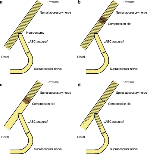
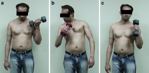
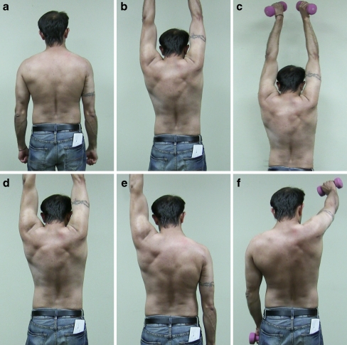
The use of end-to-side neurrorhaphy remains a controversial topic in peripheral nerve surgery. The authors report the long-term functional outcome following a modified end-to-side motor reinnervation using the spinal accessory to innervate the suprascapular nerve following a C5 to C6 avulsion injury. Additionally, functional outcomes of an end-to-end neurotization of the triceps branch to the axillary nerve and double fascicular transfer of the ulnar and medial nerve to the biceps and brachialis are presented. Excellent functional recoveries are found in respect to shoulder abduction and flexion and elbow flexion.

Electronic supplementary material: The online version of this article (doi:10.1007/s11552-009-9242-3) contains supplementary material, which is available to authorized users.


Figures

Figure 1
Figure 1
Reinnervation mechanism of an end-to-side neurotization of the suprascapular nerve to the spinal accessory nerve to restore supra/infraspinatus function and preserve trapezius function. a In this case, a neuroectomy at the coaptation site of the spinal accessory nerve was created in order to acquire reinnervation by the motor fibers. b In addition, an axonotmetic injury was created proximal to the end-to-side neurotization through a compression to induce reinnervation of the suprascapular nerve. c Wallerian degeneration proceeds distal to the axonotmetic injury following the compression. d Afterwards, axonal regeneration proceeds through the native donor and into the recipient pathway through the end-to-side repair. The LABC graft was measured at 3 cm for this case.
Figure 2
Figure 2
Restoration of elbow flexion 5.5 years following end-to-end neurotization of the ulnar nerve (flexor carpi ulnaris fascicle) to the brachialis nerve and median nerve (flexor carpi radialis fascicle) to the biceps branch of the musculocutaneous nerve. a Normal flexion on the unoperated left side is observed with a 25-lb weight. b Restoration of elbow flexion is observed on the operated right side with a 7-lb weight. A degree of pronation is also evident during this range of motion with the weight in addition to the assistance of compensatory accessory muscles. c Normal flexion while pronated is demonstrated with a 25-lb weight.
Figure 3
Figure 3
Five years and 6 months following an end-to-side neurotization of the suprascapular nerve to the spinal accessory nerve and end-to-end neurotization of the triceps branch to the axillary nerve on the right side. a At rest, the patient does not demonstrate scapular winging on the previously injured right side. b Restoration of the deltoid and supra/infraspinatus is evident with the ability to abduct the right arm to 180° without weights. In addition, hypertrophy of the lower trapezius is seen on the right side with proper scapular stabilization. c To determine the extent of these functional outcomes, the patient demonstrates proficient abduction to 180° with a 5-lb weight. d Flexion was also examined and the patient was able to flex both arms to their full range of motion of 180° without weights. e With a 5-lb weight, the patient was able to demonstrate proper flexion to 180° on the left side. f In comparison, on the right side with a 5-lb weight, the patient was only able to flex to approximately 120°.

Similar articles

Cited by

References

    1. Akeda K, et al. Regenerating axons emerge far proximal to the coaptation site in end-to-side nerve coaptation without a perineurial window using a T-shaped chamber. Plast Reconstr Surg. 2006;117:1194–1203. doi: 10.1097/01.prs.0000201460.54187.d7. - DOI - PubMed
    1. Asaoka K, Sawamura Y. Hypoglossal-facial nerve side-to-end anastomosis. J Neurosurg. 1999;91:163–164. - PubMed
    1. Asaoka K, Sawamura Y, Nagashima M, Fukushima T. Surgical anatomy for direct hypoglossal-facial nerve side-to-end "anastomosis". J Neurosurg. 1999;91:268–275. doi: 10.3171/jns.1999.91.2.0268. - DOI - PubMed
    1. Bertelli JA, Ghizoni MF. Nerve repair by end-to-side coaptation or fascicular transfer: a clinical study. J Reconstr Microsurg. 2003;19:313–318. doi: 10.1055/s-2003-42499. - DOI - PubMed
    1. Bontioti E, Kanje M, Lundborg G, Dahlin LB. End-to-side nerve repair in the upper extremity of rat. J Peripher Nerv Syst. 2005;10:58–68. doi: 10.1111/j.1085-9489.2005.10109.x. - DOI - PubMed