. 2010 Jan;51(1):181-90.
doi: 10.1002/hep.23265.

Mouse organic solute transporter alpha deficiency enhances renal excretion of bile acids and attenuates cholestasis

Affiliations

Mouse organic solute transporter alpha deficiency enhances renal excretion of bile acids and attenuates cholestasis

Carol J Soroka et al. Hepatology. 2010 Jan.

Abstract

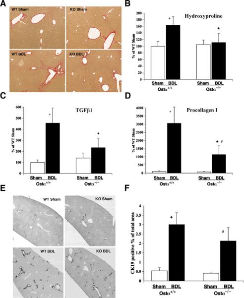
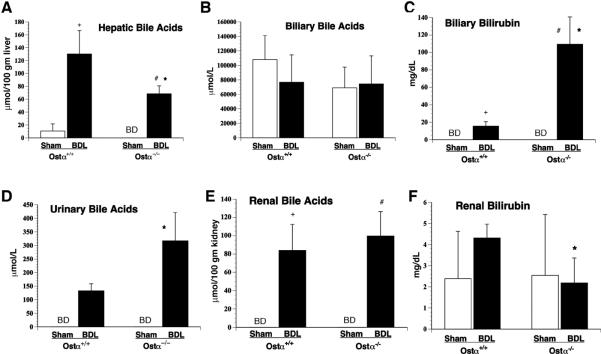
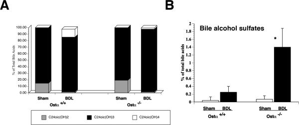
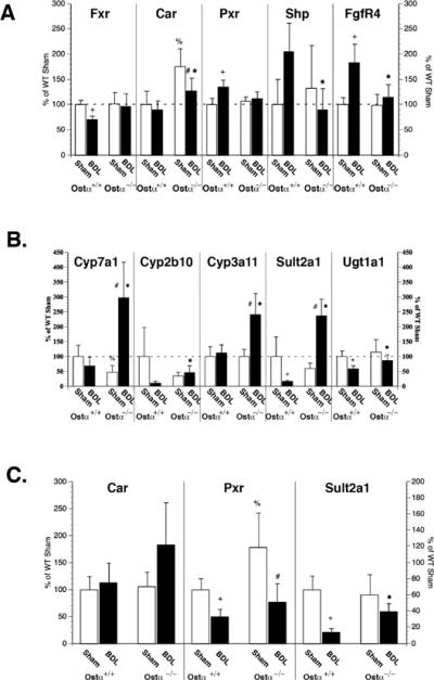
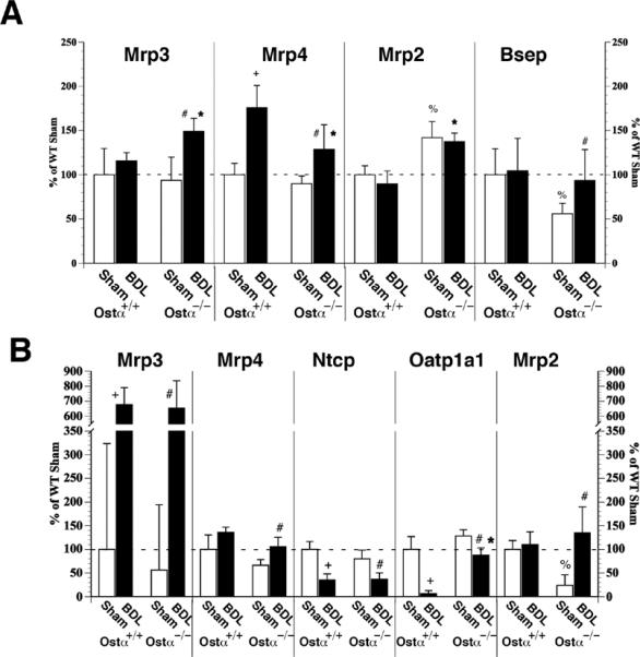
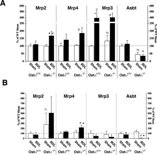
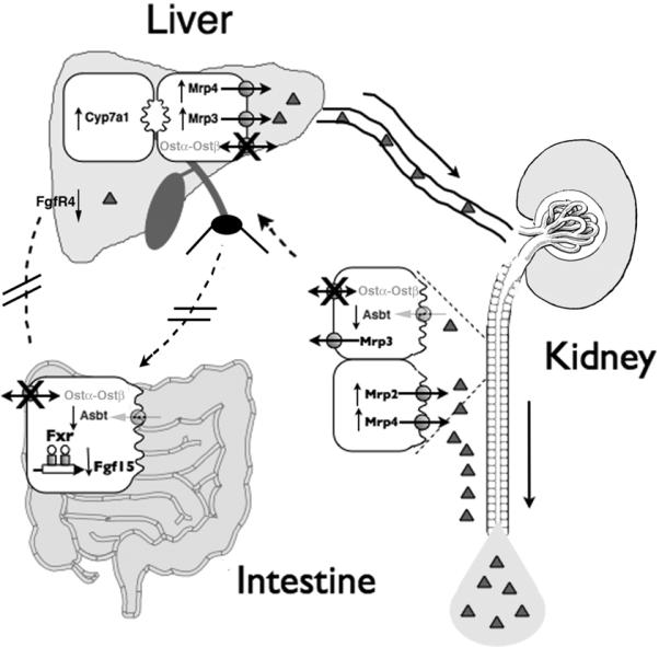
Organic solute transporter alpha-beta (Ostalpha-Ostbeta) is a heteromeric bile acid and sterol transporter that facilitates the enterohepatic and renal-hepatic circulation of bile acids. Hepatic expression of this basolateral membrane protein is increased in cholestasis, presumably to facilitate removal of toxic bile acids from the liver. In this study, we show that the cholestatic phenotype induced by common bile duct ligation (BDL) is reduced in mice genetically deficient in Ostalpha. Although Ostalpha(-/-) mice have a smaller bile acid pool size, which could explain lower serum and hepatic levels of bile acids after BDL, gallbladder bilirubin and urinary bile acid concentrations were significantly greater in Ostalpha(-/-) BDL mice, suggesting additional alternative adaptive responses. Livers of Ostalpha(-/-) mice had higher messenger RNA levels of constitutive androstane receptor (Car) than wild-type BDL mice and increased expression of Phase I enzymes (Cyp7a1, Cyp2b10, Cyp3a11), Phase II enzymes (Sult2a1, Ugt1a1), and Phase III transporters (Mrp2, Mrp3). Following BDL, the bile acid pool size increased in Ostalpha(-/-) mice and protein levels for the hepatic basolateral membrane export transporters, multidrug resistance-associated protein 3 (Mrp3) and Mrp4, and for the apical bilirubin transporter, Mrp2, were all increased. In the kidney of Ostalpha(-/-) mice after BDL, the apical bile acid uptake transporter Asbt is further reduced, whereas the apical export transporters Mrp2 and Mrp4 are increased, resulting in a significant increase in urinary bile acid excretion.

Conclusion: These findings indicate that loss of Ostalpha provides protection from liver injury in obstructive cholestasis through adaptive responses in both the kidney and liver that enhance clearance of bile acids into urine and through detoxification pathways most likely mediated by the nuclear receptor Car.


Figures

Figure 1
Figure 1
BDL results in less fibrosis in the livers of Ostα−/− mice than the Ostα+/+ controls. A. Sirius Red staining of liver sections. B. Hydroxyproline levels. 100%=157.50 mg/gm liver. C. mRNA for TGFβ1 is significantly increased in the Ostα+/+, but not Ostα−/−, mouse livers after BDL. D. mRNA levels of procollagen I are significantly lower in the Ostα−/− mice compared to the Ostα+/+ mice. E. Immunohistochemical staining for bile ducts using an antibody to cytokeratin 19. WT, wild-type; KO, knockout. F. Quantitation of cytokeratin 19 revealed less bile duct proliferation in the Ostα−/− mice, although this did not reach statistical significance (p=0.06). n=5–6; +p<0.01 Ostα+/+ Sham VS Ostα+/+ BDL; *p<0.05 Ostα+/+ BDL VS Ostα−/− BDL; #p<0.01 Ostα−/− Sham VS Ostα−/− BDL
Figure 2
Figure 2
Altered levels of hepatic and renal bile acids and bilirubin after BDL reflect adaptive changes that result in less hepatic retention and more urinary elimination of bile acids in Ostα−/− mice. A. Hepatic bile acids. B. Biliary bile acids. C. Biliary bilirubin. D. Urinary bile acids. E. Renal parenchymal bile acids. F. Renal parenchymal bilirubin. n=5–6; p<0.01, + Ostα+/+ Sham VS Ostα+/+ BDL; * Ostα+/+ BDL VS Ostα−/− BDL; # Ostα−/− Sham VS Ostα−/− BDL; BD, below detection
Figure 3
Figure 3
Analysis of liver bile acids by mass spectrometry. A. Quantification of di-, tri-, and tetra-hydroxylated bile acids was normalized to the total detectable bile acids for each animal. After BDL, Osta+/+ mice demonstrated a 35-fold increase in tetrahydroxylated bile acid which was not seen in the Osta−/− mice. B. Livers from Osta−/− mice contain significantly more bile alcohol sulfates than the Osta+/+ mice. n=5–6; *p<0.001 Ostα+/+ BDL VS Ostα−/− BDL
Figure 4
Figure 4
Gene and protein expression in livers of sham and BDL Ostα+/+ and Ostα−/− mice. A. mRNA for the nuclear receptors, Fxr and Pxr, are unchanged in Ostα−/− mice, while the xenobiotic receptor, Car, is significantly increased in sham and BDL Ostα−/− mice. Both Shp and FgfR4 mRNA are significantly increased in the Ostα+/+ BDL mice, but are lower in Ostα−/− BDL mice compared to the Ostα+/+ BDL mice. B. mRNA levels are increased for Cyp7a1, Cyp2b10, Cyp3a11, Sult2a1, Ugt1a1 in Ostα−/− BDL mice as compared to Ostα+/+ BDL mice. C. Protein expression of Car and Sult2a1, but not Pxr, tend to be elevated after BDL in Ostα−/−, but not Ostα+/+, mice. Data are normalized to SH-PTP. n=5–6; p<0.05, + Ostα+/+ Sham VS Ostα+/+ BDL; * Ostα+/+ BDL VS Ostα−/− BDL; % Ostα+/+ Sham VS Ostα−/− Sham; # Ostα−/− Sham VS Ostα−/− BDL
Figure 5
Figure 5
Membrane transporter gene and protein expression in livers of Ostα+/+ and Ostα−/− mice. A. mRNA expression. Mrp3 and Mrp4 are both higher in Ostα−/− BDL mice than the Ostα−/− sham controls. The apical transporter, Mrp2, is higher in sham and BDL Ostα−/− mice, whereas Bsep is decreased in the sham and then increased to wild-type levels after BDL. B. Protein expression. Mrp3 and Mrp4 is higher after BDL in Ostα−/− mice. Both Ntcp and Oatp1a1 are significantly decreased after BDL in the wild-type mice, but Oatp1a1 is maintained close to sham levels in the Ostα−/− mice. Protein expression of Mrp2 is down in the sham Ostα−/− mice, but increases after BDL. n=5–6; p<0.05, + Ostα+/+ Sham VS Ostα+/+ BDL; * Ostα+/+ BDL VS Ostα−/− BDL; % Ostα+/+ Sham VS Ostα−/− Sham; # Ostα−/− Sham VS Ostα−/− BDL
Figure 6
Figure 6
Membrane transporter gene and protein expression in kidneys of Ostα+/+ and Ostα−/− mice. A. mRNA expression. The apical export transporters, Mrp2 and Mrp4, are up-regulated in the kidneys of Ostα−/− mice after BDL. mRNA for the basolateral export transporter, Mrp3, is higher in the sham Ostα−/− mice than in the sham Ostα+/+ mice, but then is increased equally in the two groups after BDL. The apical bile acid uptake transporter, Asbt, is down-regulated in both the sham and BDL Ostα−/− kidneys as compared to wild-type mice. B. Protein expression. Mrp4 is significantly higher after BDL in the Ostα−/− mice. Mrp2 has a tendency to be increased in Ostα−/− mice after BDL,. There was no change in the basolateral Mrp3, whereas the apical uptake transporter, Asbt, was almost undetectable after BDL in the Ostα−/− mice. All samples were normalized to β-actin. n=5–6; p<0.05, + Ostα+/+ Sham VS Ostα+/+ BDL; * Ostα+/+ BDL VS Ostα−/− BDL; % Ostα+/+ Sham VS Ostα−/− Sham; # Ostα−/− Sham VS Ostα−/− BDL
Figure 7
Figure 7
Proposed protective mechanism in Ostα−/− mice after BDL. Ostα deficient mice have a decreased bile acid pool size and demonstrate down-regulation of the Asbt gene in both the intestine and kidney. After BDL, they up-regulate bile acid synthesis through lower levels of Shp and Fgf15, thereby increasing expression of Cyp7a1. The bile acids are transported from the liver to the circulation through an up-regulation of the basolateral transporters, Mrp3 and Mrp4, but they cannot be efficiently reabsorbed in the kidney because of the decreased expression of Asbt and the lack of Ostα-Ostβ. Furthermore, an up-regulation of the apical transporters, Mrp2 and Mrp4, efficiently prevents further accumulation of bile acids in the kidney. Therefore, ~3 fold more bile acids are excreted into the urine and removed from the body after BDL, compared to Ostα+/+ mice.

Similar articles

Cited by

References

    1. Ballatori N, Fang F, Christian WV, Li N, Hammond CL. Ostα-Ostβ is required for bile acid and conjugated steroid disposition in the intestine, kidney, and liver. Am J Physiol Gastrointest Liver Physiol. 2008;295:G179–186. - PMC - PubMed
    1. Rao A, Haywood J, Craddock AL, Belinsky MG, Kruh GD, Dawson PA. The organic solute transporter α-β, Ostα-Ostβ, is essential for intestinal bile acid transport and homeostasis. PNAS. 2008;105:3891–3896. - PMC - PubMed
    1. Ballatori N, Christian WV, Lee JY, Dawson PA, Soroka CJ, Boyer JL, Madejczyl MS, et al. OSTα-OSTβ: A Major Basolateral Bile Acid and Steroid Transporter in Human Intestinal, Renal, and Biliary Epithelia. Hepatology. 2005;42:1270–1279. - PubMed
    1. Dawson PA, Hubbert HJ, Craddock AL, Zeranque N, Christian WV, Ballatori N. The heteromeric organic solute transporter alpha-beta, Ostα-Ostβ, is an ileal basolateral bile acid transporter. J Biol Chem. 2005;280:6960–6968. - PMC - PubMed
    1. Inagaki T, Choi M, Moschetta A, Peng L, Cummins CL, McDonald JG, Luo G, et al. Fibroblast growth factor 15 functions as an enterohepatic signal to regulate bile acid homeostasis. Cell Metabolism. 2005;2:217–225. - PubMed

Publication types

Substances