. 2010 Jun;16(6):1029-39.
doi: 10.1002/ibd.21150.

Angiopoietin-2 in experimental colitis

Affiliations

Angiopoietin-2 in experimental colitis

Vijay C Ganta et al. Inflamm Bowel Dis. 2010 Jun.

Abstract

Background: The pathophysiology of inflammatory bowel disease (IBD) includes leukocyte infiltration, blood and lymphatic remodeling, weight loss and protein enteropathy. The roles of angiopoietin-2 (Ang-2) in initiating gut inflammation, leukocyte infiltration and angiogenesis are not well understood.

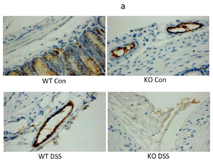
Methods: Disease activity index, histopathological scoring, myeloperoxidase assay, immunohistochemistry and sodium dodecyl sulphate- polyacrylamide gel electrophoretic methods were employed in the present study to address the roles of Ang-2 in experimental colitis.

Results: Several important differences were seen in the development of experimental IBD in Ang-2(-/-) mice. Although weight change and disease activity differ only slightly in WT and Ang-2(-/-) + DSS treated mice, leukocyte infiltration, inflammation and blood and lymphatic vessel density is significantly attenuated compared to WT + DSS mice. Gut capillary fragility and water export (stool blood and form) appear significantly earlier in Ang-2(-/-) + DSS mice vs. WT. Colon lengths were also significantly reduced in Ang-2(-/-) and gut histopathology was less severe in Ang-2(-/-) compared to WT + DSS. Lastly, the decrease in serum protein content in WT + DSS was less severe in Ang-2(-/-) + DSS, thus protein losing enteropathy (PLE) a feature of IBD is relieved by Ang-2(-/-).

Conclusion: These data demonstrate that in DSS colitis, Ang-2 mediates inflammatory hemangiogenesis, lymphangiogenesis and neutrophil infiltration to reduce some, but not all clinical features of IBD. The implications for Ang-2 manipulation in the development of IBD and other inflammatory diseases and treatments involving Ang-2 are discussed.


Figures

Figure 1
Figure 1. Disease Activity Index (DAI) and weight change
(a) Disease activity. Ang-2-/-/DSS (KO DSS) was significantly different from baseline at day 2 through day 7; WT + DSS-DAI (WT DSS) was not significantly different from baseline until day 5. (b). Weight change. Weight loss in Ang-2-/- + DSS and WT + DSS were both significantly different (*= p<0.05) from baseline at day 5 through day 7 (repeated measures ANOVA, Dunnett’s post-testing). No difference in weight was observed between. Ang-2-/-/DSS; WT + DSS.
Figure 2
Figure 2. DSS induced occult blood is exacerbated in Ang-2-/-
Occult blood was detected in the feces of Ang-2-/- + DSS treated mice as early as day 2 through day 7. WT + DSS treated mice did not show evidence of fecal blood until day 3 (through day 7). No significant presence of occult blood was seen in control (WT) or Ang-2-/- (KO Con) mice. (*= p<0.05 vs. control, ** = p<0.01 vs. control)
Figure 3
Figure 3. DSS stool form scoring is exacerbated in Ang-2-/-
Stool form scores were significantly different from baseline by day 2 in Ang-2-/- + DSS. In WT DSS mice significant alterations in stool form were observed from day 3 until day 7 (p<0.05). No significant differnce was observed in WT Con and Ang-/- Con mice.
Figure 4
Figure 4. DSS induced colon shortening is exacerbated in Ang-2-/-
Colon length in Ang-2-/- mice (7.3 +/-0.25 cm) was not significantly different from that in normal controls (7.45 +/- 0.67 cm). Colon length was significantly reduced in WT + DSS colons (to 5.56 +/- 0.13 cm) compared to WT controls (* p<0.05). Colon length was further reduced in Ang-2-/- mice (4.69 +/- 1.25 cm, **p<0.01 vs. Ang-2-/-).
Figure 5
Figure 5. Effects of Ang-2-/- status on histopathological scoring in DSS colitis
(a) Histopathology scores. WT + DSS had a cumulative histopathology score of 21.71±1.10 (Normal score=0, max injury=40), whereas, WT controls were 3±1.22 Ang-2-/-/DSS (15.54±2.06) was significantly reduced (by 28.52%) compared to WT + DSS. Ang-2-/- showed a score of 3 ±1.16. (One-way ANOVA with Bonferroni post-testing. (b). Representative images of hematoxylin/eosin stained tissue from WT Control, Ang-2 KO Con, WT DSS and Ang-2-/- + DSS treated colons.
Figure 6
Figure 6. Effects of Ang-2-/- status on blood and lymphatic vessel density scoring in DSS colitis
(a) Blood vessel (MECA-32+) immunostained vessels were significantly increased in the colon of WT + DSS (2.19 fold increase over the WT controls). (b) Similarly, lymphatic (VEGFR-3+) vessels showed a 4.9 fold increase from WT to WT + DSS. Increased lymphangiogenesis was significantly reduced in Ang-2-/- and Ang-2-/-/DSS. (c) Histogram of # vessels/section (One-way ANOVA w/Bonferroni post-testing). (Magnification = 200X).
Figure 7
Figure 7. Ang-2 regulates gut neutrophil infiltration in DSS colitis
The level of neutrophil invasion, measured by myeloperoxidase (MPO) activity of the colonic tissue, was significantly elevated in WT + DSS vs. WT, and was not significantly affect in Ang-2-/- /DSS vs. WT + DSS.
Figure 8
Figure 8. Ang-2-/- status affects serum albumin content in DSS colitis
A significant decrease in the serum protein content was observed in the WT + DSS (p<0.05 vs.controls). There was no significant loss of serum protein content in Ang-2-/- or. Ang-2-/-/DSS compared to control. (One-way ANOVA with Bonferroni post-testing).

References

    1. Ando T, Jordan P, Wang Y, et al. MAdCAM-1 expression and regulation in murine colonic endothelial cells in vitro. Inflamm Bowel Dis. 2005;11:258–264. - PubMed
    1. Bao P, Kodra A, Tomic-Canic M, et al. The role of vascular endothelial growth factor in wound healing. J Surg Res. 2009;153:347–358. - PMC - PubMed
    1. Benelli R, Albini A, Noonan D. Neutrophils and angiogenesis: potential initiators of the angiogenic cascade. Chem Immunol Allergy. 2003;83:167–181. - PubMed
    1. Benelli R, Lorusso G, Albini A, et al. Cytokines and chemokines as regulators of angiogenesis in health and disease. Curr Pharm Des. 2006;12:3101–3115. - PubMed
    1. Chidlow JH, Jr, Langston W, Greer JJ, et al. Differential angiogenic regulation of experimental colitis. Am J Pathol. 2006;169:2014–2030. - PMC - PubMed

Publication types

MeSH terms

LinkOut - more resources