Review
. 2009 Nov;5(9):1501-11.
doi: 10.2217/fon.09.109.

Clinical implications of near-infrared fluorescence imaging in cancer

Affiliations
Review

Clinical implications of near-infrared fluorescence imaging in cancer

Nobuyuki Kosaka et al. Future Oncol. 2009 Nov.

Abstract

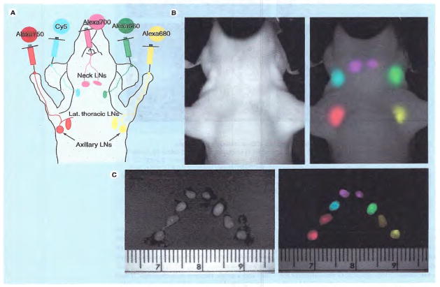
Near-infrared (NIR) fluorescence cancer imaging is a growing field for both preclinical and clinical application to the clinical management for cancer patients due to its advantageous features, including a high spatial resolution, portability, real-time display and detailed molecular profiling with the multiplexed use of fluorescent probes. In this review, we present a basic concept of NIR fluorescence imaging and overview its potential clinical applications for in vivo cancer imaging, including cancer detection/characterization, lymphatic imaging (sentinel lymph node detection) and surgical/endoscopic guidance. NIR fluorescence imaging can compensate some limitations of conventional imaging modalities, and thus it could play an important role for cancer imaging combined with other modalities in clinical practice.


Figures

Figure 1
Figure 1. Extinction coefficient of oxyhemoglobin, deoxyhemoglobin and water
The ‘diagnostic window’ is in the near-infrared range (650–900 nm) (pink), where the extinction coefficients are at their minimum.
Figure 2
Figure 2
Indocyanine green (ICG).
Figure 3
Figure 3. EGFR typing by multicolor NIR fluorescence imaging
Mouse bearing two different types of EGF receptor (EGFR)-expressed tumors, A431 overexpressing HER1 (arrows) and NIH3T3/HER2 overexpressing HER2 (arrow heads), intravenously received a cocktail of Cy5.5-labeled cetuximab (anti-HER1) and Cy7-labeled trastuzumab (anti-HER2) 24 h prior to imaging. All tumors are depicted by different spectra (absorption and emission wavelengths: 675 nm and 694 nm for Cy5.5, and 743 nm and 767 nm for Cy7, respectively). EGFR receptor expressions can be noninvasively diagnosed by its distinct colors. Note that background fluorescence (mixture of autofluorescence, unbound Cy5.5-, and Cy7-fluorescence) are eliminated by spectral unmixing algorithm [26].
Figure 4
Figure 4. In vivo cancer imaging with activatable ICG–antibody conjugates
Activatable ICG–antibody conjugate (A) is not fluorescent in phosphate buffered saline (i), while it becomes fluorescent in SDS and 2-ME added condition (ii), which can dissociate and disrupt ICG and antibody interaction. The mouse bearing ATAC4 tumor (CD25+, arrow head) and A431/RFP (CD25, arrow) intravenously received ICG-conjugated daclizumab (anti-CD25 monoclonal antibody) (B). The target tumor (ATAC4) is visualized at ICG spectrum (absorption and emission wavelengths: 780 and 820 nm, respectively), while nontarget tumor (A431/RFP) shows no fluorescence at ICG spectrum due to the quenching capability of conjugates. ICG: Indocyanine green; RFP: Red fluorescent protein; SDS: Sodium dodecyl sulfate.
Figure 5
Figure 5. Sites of injection (A), and in vivo (B) and ex vivo (C) multicolor fluorescence lymphatic images
Different lymph nodes are depicted by distinct colors. LN: Lymph node.

References

    1. Frangioni JV. New technologies for human cancer imaging. J Clin Oncol. 2008;26(24):4012–4021. Excellent review of the recent technical advances for human cancer imaging. - PMC - PubMed
    1. Brancato R, Trabucchi G. Fluorescein and indocyanine green angiography in vascular chorioretinal diseases. Semin Ophthalmol. 1998;13(4):189–198. - PubMed
    1. Flower RW, Hochheimer BF. Indocyanine green dye fluorescence and infrared absorption choroidal angiography performed simultaneously with fluorescein angiography. Johns Hopkins Med J. 1976;138(2):33–42. - PubMed
    1. Choe R, Corlu A, Lee K, et al. Diffuse optical tomography of breast cancer during neoadjuvant chemotherapy: a case study with comparison to MRI. Med Phys. 2005;32(4):1128–1139. - PubMed
    1. Demos SG, Gandour-Edwards R, Ramsamooj R, White R. Near-infrared autofluorescence imaging for detection of cancer. J Biomed Opt. 2004;9(3):587–592. - PubMed

Publication types