. 2009 Nov 10;16(1):99.
doi: 10.1186/1423-0127-16-99.

Glutamate (mGluR-5) gene expression in brain regions of streptozotocin induced diabetic rats as a function of age: role in regulation of calcium release from the pancreatic islets in vitro

Affiliations

Glutamate (mGluR-5) gene expression in brain regions of streptozotocin induced diabetic rats as a function of age: role in regulation of calcium release from the pancreatic islets in vitro

Savitha Balakrishnan et al. J Biomed Sci. .

Abstract

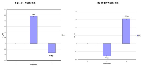
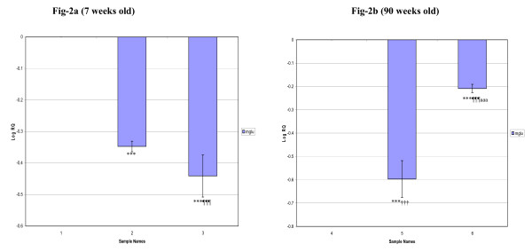
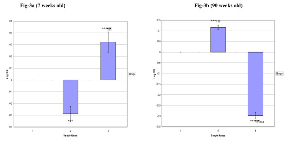
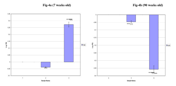
Metabotrophic glutamate receptors (mGluRs) modulate cellular activities involved in the processes of differentiation and degeneration. In this study, we have analysed the expression pattern of group-I metabotropic glutamate receptor (mGlu-5) in cerebral cortex, corpus striatum, brainstem and hippocampus of streptozotocin induced and insulin treated diabetic rats (D+I) as a function of age. Also, the functional role of glutamate receptors in intra cellular calcium release from the pancreatic islets was studied in vitro. The gene expression studies showed that mGlu-5 mRNA in the cerebral cortex increased siginficantly in 7 weeks old diabetic rats whereas decreased expression was observed in brainstem, corpus striatum and hippocampus when compared to control. 90 weeks old diabetic rats showed decreased expression in cerebral cortex, corpus striatum and hippocampus whereas in brainstem the expression increased significantly compared to their respective controls. In 7 weeks old D+I group, mGlu-5 mRNA expression was significantly decreased in cerebral cortex and corpus striatum whereas the expression increased significantly in brainstem and hippocampus. 90 weeks old D+I group showed an increased expression in cerebral cortex, while it was decreased significantly in corpus striatum, brainstem and hippocampus compared to their respective controls. In vitro studies showed that glutamate at lower concentration (10(-7) M) stimulated calcium release from the pancreatic islets. Our results suggest that mGlu-5 receptors have differential expression in brain regions of diabetes and D+I groups as a function of age. This will have clinical significance in management of degeneration in brain function and memory enhancement through glutamate receptors. Also, the regulatory role of glutamate receptors in calcium release has immense therapeutic application in insulin secretion and function.


Figures

Figure 1
Figure 1
Real-Time PCR amplification of Glutamate receptor (mGlu-5) mRNA from the Cerebral cortex of Control, Diabetic and Insulin treated Diabetic Rats. Sample Names. 1. 7 weeks Control, 2. 7 weeks Diabetic, 3. 7 weeks Insulin treated Diabetic. 4. 90 weeks Control, 5. 90 weeks Diabetic, 6. 90 weeks Insulin treated Diabetic. Real Time PCR analysis was done using mGlu-5 specific primer and fluorescently labeled Taq probe. The TaqMan reaction mixture of 20 μl contained 25 ng of total RNA-derived cDNAs, 200 nM each of the forward primer, reverse primer and TaqMan probe for glutamergic- (mGlu-5) gene, endogenous control, β-actin and 12.5 μl of TaqMan 2× Universal PCR Mastermix (Applied Biosystems). All reactions were performed in duplicates. The ΔΔCT method of relative quantification was used to determine the fold change in expression. This was done by first normalizing the resulting threshold cycle (CT) values of the target mRNAs to the CT values of the internal control β-actin in the same samples (ΔCT = CTTarget - CTβ-actin). It was further normalized with the control (ΔΔCT = ΔCT - CTControl). The fold change in expression was then obtained as (2-ΔΔCT) and expressed as log 2-ΔΔCT. Values are Mean ± S.D of 4-6 separate experiments. ***p < 0.001 when compared to control, ¶¶¶ p < 0.001 when compared to diabetic, ††† p < 0.001 when compared to 7 weeks old diabetic, aaa p < 0.001 when compared to 7 weeks old insulin treated diabetic.
Figure 2
Figure 2
Real-Time PCR amplification of Glutamate receptor (mGlu-5) mRNA from the Corpus striatum of Control, Diabetic and Insulin treated Diabetic Rats. Sample Names. 1. 7 weeks Control, 2. 7 weeks Diabetic, 3. 7 weeks Insulin treated Diabetic. 4. 90 weeks Control, 5. 90 weeks Diabetic, 6. 90 weeks Insulin treated Diabetic. Values are Mean ± S.D of 4-6 separate experiments. ***p < 0.001 when compared to control, ¶¶¶p < 0.001 when compared to diabetic, †††p < 0.001 when compared to 7 weeks old diabetic, aaap < 0.001 when compared to 7 weeks old insulin treated diabetic. Other details as in the legend to Figure-1.
Figure 3
Figure 3
Real-Time PCR amplification of Glutamate receptor (mGlu-5) mRNA from the Brainstem of Control, Diabetic and Insulin treated Diabetic Rats. Sample Names. 1. 7 weeks Control, 2. 7 weeks Diabetic, 3. 7 weeks Insulin treated Diabetic. 4. 90 weeks Control, 5. 90 weeks Diabetic, 6. 90 weeks Insulin treated Diabetic. Values are Mean ± S.D of 4-6 separate experiments. ***p < 0.001 when compared to control, ¶¶¶p < 0.001 when compared to diabetic, †††p < 0.001 when compared to 7 weeks old diabetic, aaap < 0.001 when compared to 7 weeks old insulin treated diabetic. Other details as in the legend to Figure-1.
Figure 4
Figure 4
Real-Time PCR amplification of Glutamate receptor (mGlu-5) mRNA from the Hippocampus of Control, Diabetic and Insulin treated Diabetic Rats. Sample Names. 1. 7 weeks Control, 2. 7 weeks Diabetic, 3. 7 weeks Insulin treated Diabetic. 4. 90 weeks Control, 5. 90 weeks Diabetic, 6. 90 weeks Insulin treated Diabetic. Values are Mean ± S.D of 4-6 separate experiments. ***p < 0.001 when compared to control, ¶¶¶p < 0.001 when compared to diabetic, †††p < 0.001 when compared to 7 weeks old diabetic, aaap < 0.001 when compared to 7 weeks old insulin treated diabetic. Other details as in the legend to Figure-1.
Figure 5
Figure 5
The isolated pancreatic islets were incubated for 4 hours at room temperature in 1 ml of RPMI medium containing 5 μM of Ca2+ fluorescent dye, fluo 4-AM. The images were continuously acquired before and after addition of 10-7 M of glutamate, at time intervals of 1.318 seconds, for a total of 600 seconds. Time series experiments were performed collecting 512 × 512 pixel images at 400 Hz. Fluorescence intensity was analysed using the quantitation mode in LAS-AF software from Leica Microsystems, Germany. A region of interest (ROI) was drawn within a field of view. The pixel intensity was calculated for each image in the 600 seconds sequence to analyse the intracellular Ca2+ release from the pancreatic islet cells in experimental conditions.
Figure 6
Figure 6
The isolated pancreatic islets were incubated for 4 hours at room temperature in 1 ml of RPMI medium containing 5 μM of Ca2+ fluorescent dye, fluo 4-AM. The images were continuously acquired before and after addition of 10-4 M of glutamate, at time intervals of 1.318 seconds, for a total of 600 seconds. Time series experiments were performed collecting 512 × 512 pixel images at 400 Hz. Fluorescence intensity was analysed using the quantitation mode in LAS-AF software from Leica Microsystems, Germany. A region of interest (ROI) was drawn within a field of view. The pixel intensity was calculated for each image in the 600 seconds sequence to analyse the intracellular Ca2+ release from the pancreatic islet cells in experimental conditions.

Similar articles

Cited by

References

    1. Alvarez FJ, Dewey DE, Carr PA, Cope TC, Fyffe RE. Downregulation of metabotropic glutamate receptor 1a in motoneurons after axotomy. Neuroreport. 1997;8:1711–1716. doi: 10.1097/00001756-199705060-00029. - DOI - PubMed
    1. Pin JP, Duvoisin R. The metabotropic glutamate receptors: structure and functions. Neuropharmacology. 1995;34:1–26. doi: 10.1016/0028-3908(94)00129-G. - DOI - PubMed
    1. Hollmann M, Heinemann S. Cloned glutamate receptors. Annu Rev Neurosci. 1994;17:31–108. doi: 10.1146/annurev.ne.17.030194.000335. - DOI - PubMed
    1. Lourenco Neto F, Schadrack J, Berthele A, Zieglgansberger W, Tolle TR, Castro-Lopes JM. Differential distribution of met;abotropic glutamate receptor subtype mRNAs in the thalamus of the rat. Brain Res. 2000;854:93–105. doi: 10.1016/S0006-8993(99)02326-4. - DOI - PubMed
    1. Masu M, Tanabe Y, Tsuchida K, Shigemoto R, Nakanishi S. Sequence and expression of a metabotropic glutamate receptor. Nature. 1991;349:760–765. doi: 10.1038/349760a0. - DOI - PubMed

Publication types