

Assessing the usefulness of a novel MRI-based breast density estimation algorithm in a cohort of women at high genetic risk of breast cancer: the UK MARIBS study
- PMID: 19903338
- PMCID: PMC2815542
- DOI: 10.1186/bcr2447
Assessing the usefulness of a novel MRI-based breast density estimation algorithm in a cohort of women at high genetic risk of breast cancer: the UK MARIBS study
Abstract
Introduction: Mammographic breast density is one of the strongest known risk factors for breast cancer. We present a novel technique for estimating breast density based on 3D T1-weighted Magnetic Resonance Imaging (MRI) and evaluate its performance, including for breast cancer risk prediction, relative to two standard mammographic density-estimation methods.
Methods: The analyses were based on MRI (n = 655) and mammography (n = 607) images obtained in the course of the UK multicentre magnetic resonance imaging breast screening (MARIBS) study of asymptomatic women aged 31 to 49 years who were at high genetic risk of breast cancer. The MRI percent and absolute dense volumes were estimated using our novel algorithm (MRIBview) while mammographic percent and absolute dense area were estimated using the Cumulus thresholding algorithm and also using a 21-point Visual Assessment scale for one medio-lateral oblique image per woman. We assessed the relationships of the MRI and mammographic measures to one another, to standard anthropometric and hormonal factors, to BRCA1/2 genetic status, and to breast cancer risk (60 cases) using linear and Poisson regression.
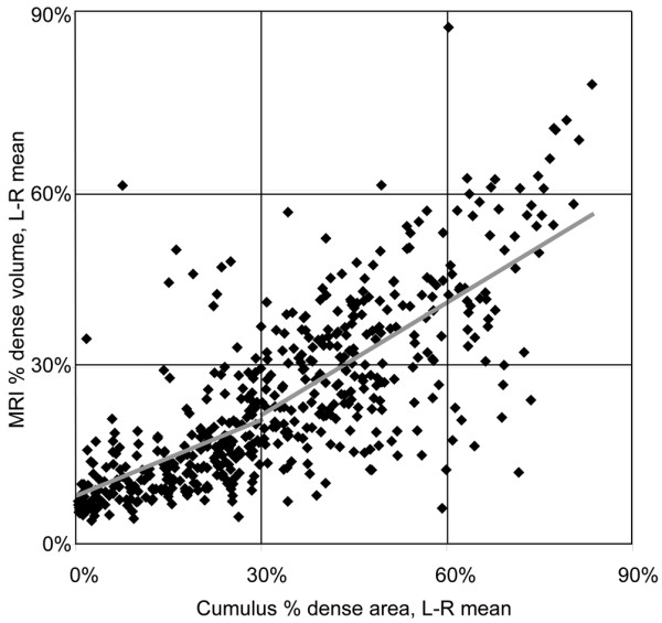
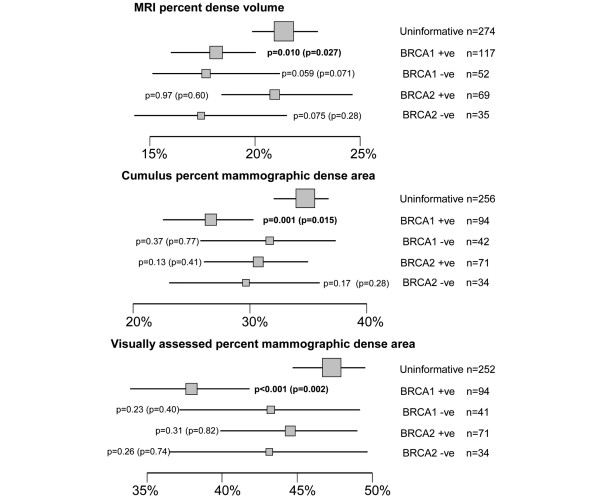
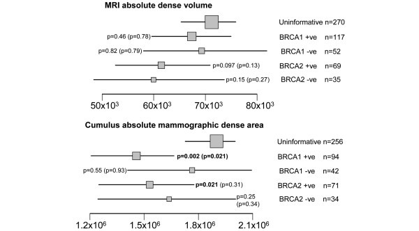
Results: MRI percent dense volume is well correlated with mammographic percent dense area (R = 0.76) but overall gives estimates 8.1 percentage points lower (P < 0.0001). Both show strong associations with established anthropometric and hormonal factors. Mammographic percent dense area, and to a lesser extent MRI percent dense volume were lower in BRCA1 carriers (P = 0.001, P = 0.010 respectively) but there was no association with BRCA2 carrier status. The study was underpowered to detect expected associations between percent density and breast cancer, but women with absolute MRI dense volume in the upper half of the distribution had double the risk of those in the lower half (P = 0.009).
Conclusions: The MRIBview estimates of volumetric breast density are highly correlated with mammographic dense area but are not equivalent measures; the MRI absolute dense volume shows potential as a predictor of breast cancer risk that merits further investigation.
Figures
Similar articles
-
Predictors of mammographic density among women with a strong family history of breast cancer.BMC Cancer. 2019 Jun 26;19(1):631. doi: 10.1186/s12885-019-5855-2. BMC Cancer. 2019. PMID: 31242899 Free PMC article.
-
Screening with magnetic resonance imaging and mammography of a UK population at high familial risk of breast cancer: a prospective multicentre cohort study (MARIBS).Lancet. 2005 May 21-27;365(9473):1769-78. doi: 10.1016/S0140-6736(05)66481-1. Lancet. 2005. PMID: 15910949
-
A pilot study of compositional analysis of the breast and estimation of breast mammographic density using three-dimensional T1-weighted magnetic resonance imaging.Cancer Epidemiol Biomarkers Prev. 2008 Sep;17(9):2268-74. doi: 10.1158/1055-9965.EPI-07-2547. Cancer Epidemiol Biomarkers Prev. 2008. PMID: 18768492 Free PMC article.
-
Supplemental Screening for Breast Cancer in Women With Dense Breasts: A Systematic Review for the U.S. Preventive Service Task Force [Internet].Rockville (MD): Agency for Healthcare Research and Quality (US); 2016 Jan. Report No.: 14-05201-EF-3. Rockville (MD): Agency for Healthcare Research and Quality (US); 2016 Jan. Report No.: 14-05201-EF-3. PMID: 26866210 Free Books & Documents. Review.
-
Breast cancer and ovarian cancer genetics.J Long Term Eff Med Implants. 2005;15(5):533-45. doi: 10.1615/jlongtermeffmedimplants.v15.i5.60. J Long Term Eff Med Implants. 2005. PMID: 16218901 Review.
Cited by
-
Impact of positional difference on the measurement of breast density using MRI.Med Phys. 2015 May;42(5):2268-75. doi: 10.1118/1.4917083. Med Phys. 2015. PMID: 25979021 Free PMC article.
-
Comparison of breast tissue measurements using magnetic resonance imaging, digital mammography and a mathematical algorithm.Phys Med Biol. 2012 Nov 7;57(21):6903-27. doi: 10.1088/0031-9155/57/21/6903. Epub 2012 Oct 9. Phys Med Biol. 2012. PMID: 23044556 Free PMC article.
-
MRI background parenchymal enhancement, breast density and breast cancer risk factors: A cross-sectional study in pre- and post-menopausal women.NPJ Breast Cancer. 2022 Aug 25;8(1):97. doi: 10.1038/s41523-022-00458-2. NPJ Breast Cancer. 2022. PMID: 36008488 Free PMC article.
-
Effect of taxane-based neoadjuvant chemotherapy on fibroglandular tissue volume and percent breast density in the contralateral normal breast evaluated by 3T MR.NMR Biomed. 2013 Dec;26(12):1705-13. doi: 10.1002/nbm.3006. Epub 2013 Aug 12. NMR Biomed. 2013. PMID: 23940080 Free PMC article.
-
Consistency of breast density measured from the same women using different MR scanners.Ann Oncol. 2011 Dec;22(12):2693-2694. doi: 10.1093/annonc/mdr456. Epub 2011 Oct 19. Ann Oncol. 2011. PMID: 22015449 Free PMC article. No abstract available.
References
-
- Mitchell G, Antoniou AC, Warren R, Peock S, Brown J, Davies R, Mattison J, Cook M, Warsi I, Evans DG, Eccles D, Douglas F, Paterson J, Hodgson S, Izatt L, Cole T, Burgess L, Eeles R, Easton DF. Mammographic density and breast cancer risk in BRCA1 and BRCA2 mutation carriers. Cancer Res. 2006;66:1866–1872. doi: 10.1158/0008-5472.CAN-05-3368. - DOI - PubMed
-
- Khazen M, Warren RM, Boggis CR, Bryant EC, Reed S, Warsi I, Pointon LJ, Kwan-Lim GE, Thompson D, Eeles R, Easton D, Evans DG, Leach MO. A pilot study of compositional analysis of the breast and estimation of breast mammographic density using three-dimensional T1-weighted magnetic resonance imaging. Cancer Epidemiol Biomarkers Prev. 2008;17:2268–2274. doi: 10.1158/1055-9965.EPI-07-2547. - DOI - PMC - PubMed
-
- Brown J, Buckley D, Coulthard A, Dixon AK, Dixon JM, Easton DF, Eeles RA, Evans DG, Gilbert FG, Graves M, Hayes C, Jenkins JP, Jones AP, Keevil SF, Leach MO, Liney GP, Moss SM, Padhani AR, Parker GJ, Pointon LJ, Ponder BA, Redpath TW, Sloane JP, Turnbull LW, Walker LG, Warren RM. Magnetic resonance imaging screening in women at genetic risk of breast cancer: imaging and analysis protocol for the UK multicentre study. UK MRI Breast Screening Study Advisory Group. Magn Reson Imaging. 2000;18:765–776. doi: 10.1016/S0730-725X(00)00167-3. - DOI - PubMed
-
- Leach MO, Boggis CR, Dixon AK, Easton DF, Eeles RA, Evans DG, Gilbert FJ, Griebsch I, Hoff RJ, Kessar P, Lakhani SR, Moss SM, Nerurkar A, Padhani AR, Pointon LJ, Thompson D, Warren RM. Screening with magnetic resonance imaging and mammography of a UK population at high familial risk of breast cancer: a prospective multicentre cohort study (MARIBS) Lancet. 2005;365:1769–1778. doi: 10.1016/S0140-6736(05)66646-9. - DOI - PubMed
Publication types
MeSH terms
Grants and funding
LinkOut - more resources
Full Text Sources
Medical
Miscellaneous
