. 2009 Nov 10:8:11.
doi: 10.1186/1477-5751-8-11.

Human spongiosa mesenchymal stem cells fail to generate cardiomyocytes in vitro

Affiliations

Human spongiosa mesenchymal stem cells fail to generate cardiomyocytes in vitro

Svetlana Mastitskaya et al. J Negat Results Biomed. .

Abstract

Background: Human mesenchymal stem cells (hMSCs) are broadly discussed as a promising cell population amongst others for regenerative therapy of ischemic heart disease and its consequences. Although cardiac-specific differentiation of hMSCs was reported in several in vitro studies, these results were sometimes controversial and not reproducible.

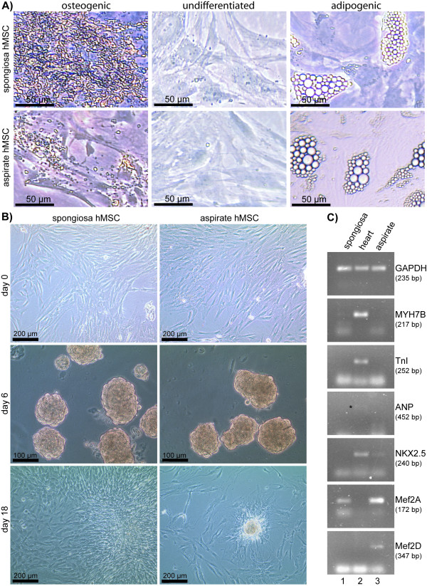
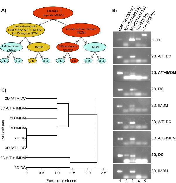
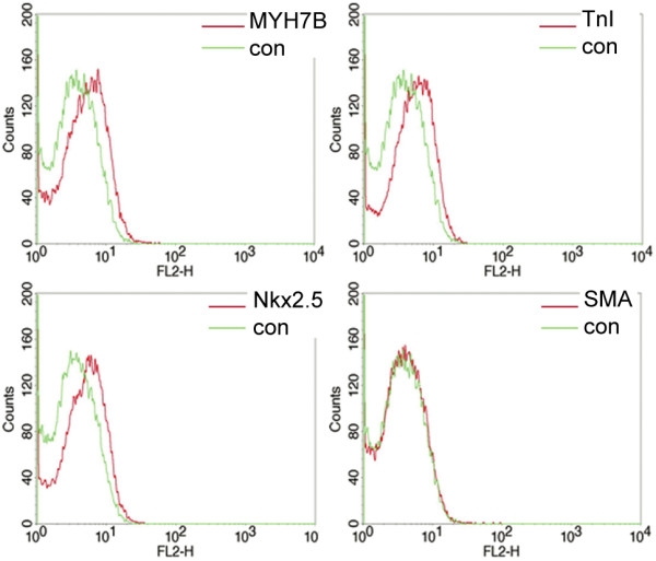
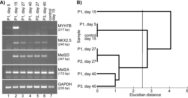
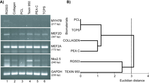
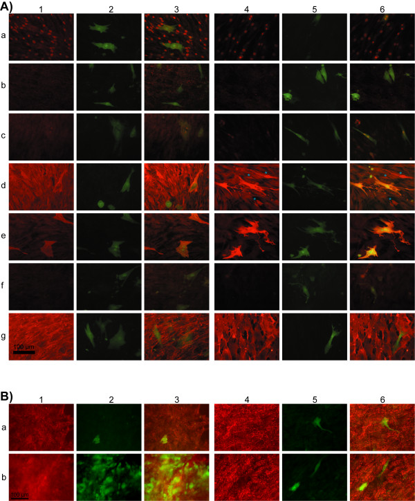
Results: In our study we have analyzed different published protocols of cardiac differentiation of hMSCs and their modifications, including the use of differentiation cocktails, different biomaterial scaffolds, co-culture techniques, and two- and three-dimensional cultures. We also studied whether 5'-azacytidin and trichostatin A treatments in combination with the techniques mentioned above can increase the cardiomyogenic potential of hMSCs. We found that hMSCs failed to generate functionally active cardiomyocytes in vitro, although part of the cells demonstrated increased levels of cardiac-specific gene expression when treated with differentiation factors, chemical substances, or co-cultured with native cardiomyocytes.

Conclusion: The failure of hMSCs to form cardiomyocytes makes doubtful the possibility of their use for mechanical reparation of the heart muscle.


Figures

Figure 1
Figure 1
Spongiosa and aspirate hMSCs cultures. (A) Spongiosa and aspirate hMSCs were cultured in stem cell medium alone (undifferentiated) or in culture medium supplemented with stimuli inducing osteogenic differentiation (osteogenic) and adipogenic differentiation (adipogenic), respectively. In both, spongiosa hMSCs and aspirate hMSCs, mineralization nodules formation confirmed osteogenic differentiation and adipogenic differentiation was confirmed by lipid vacuols. (B) Figure shows the photographs of the 3-D hMSCs cultures in differentiation cocktail on two different time points as well as 2-D cultures on starting point of the experiment (day 0). Day 6: spongiosa and aspirate hMSCs cultured on nonadhesive Petri plastic dishes on the day 6 by the method of hanging drops in differentiation cocktail. Day 18: the same cells transferred onto tissue culture plastic; the spongiosa hMSCs culture almost reached 100% confluence while aspirate hMSCs still keep together in the form of bodies and almost do not proliferate. (C) PCR from undifferentiated aspirate and spongiosa hMSCs and human adult heart tissue demonstrating the expression of Nkx2.5, MEF2A, and MEF2D genes in undifferentiated aspirate hMSCs and MEF2A and MYH7B in untreated spongiosa hMSCs. The expression of MEF2A and MEF2D genes wasn't revealed and ANP gene expression was negligible in adult human heart tissue (the band marked with black star).
Figure 2
Figure 2
Test of culture conditions for cardiac differentiation of hMSCs. (A) Scheme of experiment on cardiac differentiation of aspirate hMSCs in vitro. (B) PCR from human heart tissue and aspirate hMSCs used in 8 differentiation protocols (see A) demonstrating the highest efficacy of two of them that led to the increase in expression of Nkx2.5 gene along with MYH7B gene expression: 3D, DC and 2D, A/T+IMDM. (C) Clusterization of cell cultures based on the level of MYH7B, Nkx2.5, MEF2A, and MEF2D gene expression. The cell cultures tested in this experiment formed two statistically different (P = 0.036, ANOSIM) clusters at Euclidian distance of about 2.2. Note that the threedimensional cell culture in DC (3D DC) and the two-dimensional culture in IMDM pretreated with 5-azacytidine and trichostatin A (2D A/T + IMDM) formed a separate cluster. (2D - two-dimensional culture; 3D - three-dimensional culture; A/T - pretreatment with 5-azacytidin and trichostatin A; DC - cells cultured in differentiation cocktail; NCM - normal culture medium; IMDM - NCM based on Iscove's Modified Dulbecco's Medium; Differentiation cocktail - NCM based on DMEM-LG with components of differentiation cocktail).
Figure 3
Figure 3
Expression of cardiac-specific markers by late passage (P3) spongiosa hMSCs cultured in 3-D culture with differentiation cocktail. FACS analyses of differentiated spongiosa hMSCs stained by antibodies against cardiac myosin heavy chain (MYH7B), cardiac troponin I (TnI), Nkx2.5 and smooth muscle actin (SMA). The slight expression of MYH7B, TnI and Nkx2.5 by all cells and SMA expression by some single cells was revealed.
Figure 4
Figure 4
Cardiac-specific differentiation of spongiosa hMSC cultured 3-D in differentiation cocktail on different time points (passaged cells as well as longterm culture). (A) PCR from differentiated spongiosa hMSCs demonstrating the temporary pattern of cardiac specific gene expression. The expression of MYH7B and Nkx2.5 was high by the day 15 and then levelled off both in long-term culture and passaged cells. (B) Clusterization of cell cultures based on the level of MYH7B, Nkx2.5, MEF2A, and MEF2D gene expression on different time points and passages. The cell cultures tested in this experiment formed three statistically different (P = 0.01, ANOSIM) clusters at Euclidian distance of about 2.5. Note that one of the clusters is formed by first passage MSCs cultured in differentiation cocktail for 15 days (P1, day 15).
Figure 5
Figure 5
Expression of cardiac-specific genes by spongiosa hMSCs cultured in DC on biomaterials. (A) PCR from spongiosa hMSCs cultured on biomaterials demonstrating the effectiveness of RG503 and Texin 950 as scaffolds for supporting of cardiac differentiation of hMSCs in vitro. (B) Clusterization of cell cultures grown on different biomaterials based on the level of MYH7B, Nkx2.5, MEF2A, and MEF2D gene expression. The cell cultures tested in this experiment formed two clusters at Euclidian distance of about 2.8. The level of dissimilarity between these clusters was found to be at the edge of statistical significance (P = 0.06, ANOSIM). Note that the cells cultured on biomaterials RG503 and Texin 950 formed a separate cluster. (TCPS = tissue culture polystyrene).
Figure 6
Figure 6
Expression of myocyte-specific markers by hMSCs co-cultured with Cor.AT cells. (A) 14 days co-culture (1-3) Co-culture in CorAT medium supplemented with components of differentiation cocktail, (4-6) co-culture in Cor.AT medium: 1 and 4 - immunofluorescent staining; 2 and 5 - same field, green coloured cells - murine atrial-like cardiomyocytes (Cor.AT cells) expressing GFP; 3 and 6 - merged. (a) GATA4, (b) connexin-43, (c) Nkx2.5, (d) myoglobin (differentiated hMSCs showing organized myoglobin structure are marked with a blue star), (e) cardiac troponin I, (f) cardiac myosin heavy chain, (g) CD44. Scale bar is 100 μm. (B) Detection of connexin-43 in 25 days co-culture (1-3) Co-culture in CorAT medium supplemented with components of differentiation cocktail, (4-6) co-culture in Cor.AT medium: 1 and 4 - immunofluorescent staining; 2 and 5 - same field, green coloured cells - murine atrial-like cardiomyocytes (Cor.AT cells) expressing GFP; 3 and 6 - merged. (a) and (b) detection of connexin-43 in two independent assays after 25 days. Scale bar is 100 μm.

Similar articles

Cited by

References

    1. Brehm M, Zeus T, Strauer BE. Stem cells - clinical application and perspectives. Herz. 2002;27:611–620. doi: 10.1007/s00059-002-2435-y. - DOI - PubMed
    1. Pittenger MF, Mackay AM, Beck SC, Jaiswal RK, Douglas R, Mosca JD, Moorman MA, Simonetti DW, Craig S, Marshak DR. Multilineage potential of adult human mesenchymal stem cells. Science. 1999;284:143–146. doi: 10.1126/science.284.5411.143. - DOI - PubMed
    1. Bittira B, Kuang J-Q, Al-Khaldi A, Shum-Tim D, Chiu RC-J. In vitro preprogramming of marrow stromal cells for myocardial regeneration. Ann Thorac Surg. 2002;74:1154–1160. doi: 10.1016/S0003-4975(02)03857-2. - DOI - PubMed
    1. Gojo S, Gojo N, Takeda Y, Mori T, Abe H, Kyo S, Hata J-I, Umezawa A. In vivo cardiovasculogenesis by direct injection of isolated adult mesenchymal stem cells. Exp Cell Res. 2003;288:51–59. doi: 10.1016/S0014-4827(03)00132-0. - DOI - PubMed
    1. Mackenzie TS, Flake AW. Multilineage differentiation of human MSC after in utero transplantation. Cytotherapy. 2001;3:403–405. doi: 10.1080/146532401753277571. - DOI - PubMed

Publication types

LinkOut - more resources