. 2009 Nov 10:9:130.
doi: 10.1186/1471-2229-9-130.

Vitamin B6 deficient plants display increased sensitivity to high light and photo-oxidative stress

Affiliations

Vitamin B6 deficient plants display increased sensitivity to high light and photo-oxidative stress

Michel Havaux et al. BMC Plant Biol. .

Abstract

Background: Vitamin B6 is a collective term for a group of six interconvertible compounds: pyridoxine, pyridoxal, pyridoxamine and their phosphorylated derivatives. Vitamin B6 plays essential roles as a cofactor in a range of biochemical reactions. In addition, vitamin B6 is able to quench reactive oxygen species in vitro, and exogenously applied vitamin B6 protects plant cells against cell death induced by singlet oxygen (1O2). These results raise the important question as to whether plants employ vitamin B6 as an antioxidant to protect themselves against reactive oxygen species.

Results: The pdx1.3 mutation affects the vitamin B6 biosynthesis enzyme, pyridoxal synthase (PDX1), and leads to a reduction of the vitamin B6 concentration in Arabidopsis thaliana leaves. Although leaves of the pdx1.3 Arabidopsis mutant contained less chlorophyll than wild-type leaves, we found that vitamin B6 deficiency did not significantly impact photosynthetic performance or shoot and root growth. Chlorophyll loss was associated with an increase in the chlorophyll a/b ratio and a selective decrease in the abundance of several PSII antenna proteins (Lhcb1/2, Lhcb6). These changes were strongly dependent on light intensity, with high light amplifying the difference between pdx1.3 and the wild type. When leaf discs were exposed to exogenous 1O2, lipid peroxidation in pdx1.3 was increased relative to the wild type; this effect was not observed with superoxide or hydrogen peroxide. When leaf discs or whole plants were exposed to excess light energy, 1O2-mediated lipid peroxidation was enhanced in leaves of the pdx1.3 mutant relative to the wild type. High light also caused an increased level of 1O2 in vitamin B6-deficient leaves. Combining the pdx1.3 mutation with mutations affecting the level of 'classical' quenchers of 1O2 (zeaxanthin, tocopherols) resulted in a highly photosensitive phenotype.

Conclusion: This study demonstrates that vitamin B6 has a function in the in vivo antioxidant defense of plants. Thus, the antioxidant activity of vitamin B6 inferred from in vitro studies is confirmed in planta. Together with the finding that chloroplasts contain vitamin B6 compounds, the data show that vitamin B6 functions as a photoprotector that limits 1O2 accumulation in high light and prevents 1O2-mediated oxidative damage.


Figures
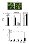
Figure 1
Figure 1
Pigment content of young leaves of WT Arabidopsis and of the pdx1 mutant. A) Plants aged 4 weeks. B) Chlorophyll and carotenoid content of young leaves. Chl, total chlorophyll; Xanth, xanthophylls; β-car, β-carotene. C) Level of various chlorophyll precursors in young leaves: Pchlide, protochlorophyllide; Chlide, chlorophyllide; GG-, DHGG- and THGG-Chl, geranylgeranyl-chlorophyll, dihydrogeranylgeranyl-chlorophyll and tetrahydrogeranylgeranyl-chlorophyll, respectively. Data are mean values of 4 measurements + SD. *, significantly different from the WT value with P < 0.01 (t test).
Figure 2
Figure 2
A) Chlorophyll content and B) chlorophyll a/b ratio in leaves of WT and pdx1 plants grown at different PFDs. Data are mean values of 3 measurements ± SD.
Figure 3
Figure 3
Photosynthetic parameters of WT Arabidopsis leaves and leaves of the pdx1 mutant grown under control conditions (150-200 μmol m-2 s-1, 25°C). A) Quantum yield of PSII photochemistry (ΔF/Fm'), B) oxygen exchange and C) NPQ measured at different PFDs. Data are mean values of 3 or 4 measurements ± SD. D) Light-induced conversion of violaxanthin (V) into zeaxanthin (Z) and antheraxanthin (A), as calculated by the equation (A+Z)/(V+A+Z). Zeaxanthin synthesis was induced by white light of PFD 1000 μmol m-2 s-1. Each point corresponds to a different leaf (1 measurement per point).
Figure 4
Figure 4
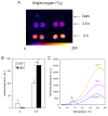
Oxidative stress in Arabidopsis leaf discs (WT and pdx1) exposed to the 1O2 generator eosin (0.5%). A) Autoluminescence imaging of leaf discs exposed for 3.5 h or 5 h to eosin in the light (400 μmol photons m-2 s-1). 'Dark' corresponds to eosin-infiltrated leaf discs kept in the dark for 5 h. B) Autoluminescence intensity in leaf discs exposed for 0 or 5 h to eosin in the light. Data are mean values of 10 measurements + SD. *, significantly different from the WT value with P < 0.001 (t test). C) Thermoluminescence band at high temperature (ca. 135°C) in leaf discs exposed for 5 h to eosin in the light. Control, leaf discs from pdx1 kept in eosin in the dark. Control WT disks (not shown) was in the same thermoluminescence intensity range. The band peaking at ca. 60°C in the control is typical of Arabidopsis. Its origin is unknown; it is not related to lipid peroxidation and could be due to thermolysis of a (yet unidentified) volatile compound [84].
Figure 5
Figure 5
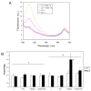
Photooxidative stress in leaf discs (WT and pdx1). A) Autoluminescence of leaf discs exposed for 6 h to 1500 μmol m-2 s-1 at 10°C. B) Thermoluminescence band at high temperature (ca. 135°C) in leaf discs exposed to high light stress for 0, 5, 6 or 20 h. The thermoluminescence signal of discs taken from leaves of the pdx1 mutant and preinfiltrated with vitamin B6 (2 mM) is also shown (5 h + vitamin B6).
Figure 6
Figure 6
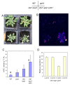
Photooxidative stress of whole Arabidopsis plants (WT and pdx1). A) Autoluminescence imaging of lipid peroxidation after high light stress (2d, 6°C, 1500 μmol m-2 s-1). B) Thermoluminescence signal of WT leaves and leaves of the pdx1 mutant before and after high light stress (LL and HL, respectively). C) Lipid hydroperoxide level (HOTE) in leaves of control and high light-stressed WT and pdx1 plants. *, significantly different from the WT value with P < 0.015 (t test). D) Distribution of lipid hydroperoxide (HOTE) isomers in leaves of control and high-light stressed WT and pdx1 plants. Data are mean values of 3 to 5 measurements + SD.
Figure 7
Figure 7
Fluorescence of SOGS in WT and mutant (pdx1) leaves exposed to high light. A) Fluorescence of leaves infiltrated with SOGS after exposure to white light (HL = 450 μmol photon m-2 s-1 for 40 min). Controls (= c) were kept in dim light before fluorescence measurements. B) Fluorescence ratio F525/F680 of WT leaves and mutant leaves infiltrated with SOGS and/or vitamin B6 before or after illumination. Data are mean values of 3 measurements + SD. *, significantly different from the WT value with P < 0.025 (t test).
Figure 8
Figure 8
Effects of high light stress (1000 μmol photons m-2 s-1 at 10°C for 2 d) on WT plants and on pdx1, vte1 npq1 and vte1 npq1 pdx1 mutant plants. A) Plants after the high light treatment. B) Autoluminescence imaging of lipid peroxidation. C) HOTE level. a, significantly different with P < 0.03 (t test). D) Distribution of HOTE isomers in leaves of the vte1 npq1 pdx1 triple mutant exposed to the high light treatment. Data are mean values of 3 or 4 measurements + SD.
Figure 9
Figure 9
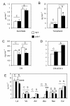
Levels of chlorophyll and various antioxidants in WT leaves and leaves of pdx1 after long-term exposure to high light (1000 μmol m-2 s-1, 10°C, 7d). A) Ascorbate, B) α-Tocopherol, C) Total chlorophyll, D) Chlorophyll a/b ratio, E) β-carotene (car) and xanthophylls (lutein (lut), violaxanthin (vio), antheraxanthin (ant), zeaxanthin (zea), neoxanthin (neo)). Data are mean values of 3 measurements + SD. C = control plants; S = plants exposed to the high light treatment. *, ** and ***, significantly different from the WT value with P < 0.001, 0.035 and 0.01, respectively (t test). White bars, WT; black bars, pdx1 mutant.
Figure 10
Figure 10
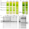
A) Separation of pigmented photosynthetic complexes of thylakoids prepared from leaves of WT and pdx1 by solubilization in 0.8% dodecylmaltoside and ultracentrifugation on sucrose gradient. Thylakoids were prepared from leaves of WT and pdx1 grown in low light (c, 200 μmol photons m-2 s-1) or acclimated for 7 d to high light (hl, 1000 μmol m-2 s-1). B1, free pigments; B2, monomeric Lhcb antennae; B3, LHCII trimers; B5, PSII core (monomeric), B6, PSI-LHCI supercomplex. The B4 band (LHCII-CP29-CP24 supercomplex, see [85]) is not visible in this gradient. B) Ultracentrifugation gradient of thylakoids (pdx1, c and hl) solubilized in 1.2% dodecylmaltoside. In the control pdx1 sample, an additional band appeared in the bottom of the gradient, which was hardly visible at 0.8% dodecylmaltoside and which corresponded to dimeric PSI-LHCI. This is presumably due to an artificial aggregation the high detergent concentration used in this preparation as previously found [86]; the same phenomenon was observed with WT thylakoids (data not shown). C and D) SDS-PAGE separation of the B2, B3 and B6 bands using two different buffer systems: tricine (C) and urea (D). See ref. [87] for identification of the bands. BBY = PSII-enriched membranes used as a reference for the PSII proteins.
Figure 11
Figure 11
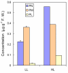
Vitamin B6 components (expressed in μg/g fresh weight) in leaves of Arabidopsis plants grown in low light (LL) or acclimated for 7 d to high light (HL, 1000 μmol photons m-2 s-1 at 10°C). F. W. = fresh weight. PM, pyridoxamine; PN, pyridoxine; PL, pyridoxal. Data are mean values of 2 or 3 measurements + SD.

Similar articles

Cited by

References

    1. Drewke C, Leistner E. Biosynthesis of vitamin B6 and structurally related derivatives. Vitam Horm. 2001;61:121–155. - PubMed
    1. Roje S. Vitamin B biosynthesis in plants. Phytochemistry. 2007;68:1904–1921. - PubMed
    1. Mooney S, Leuendorf JE, Hendrickson C, Hellmann H. Vitamin B6: a long known compound of surprising complexity. Molecules. 2009;14:329–351. - PMC - PubMed
    1. Tambasco-Studart M, Titiz O, Raschle T, Forster G, Amrhein N, Fitzpatrick TB. Vitamin B6 biosynthesis in higher plants. Proc Natl Acad Sci USA. 2005;102:13687–13692. - PMC - PubMed
    1. Tambasco-Studart M, Tews I, Amrhein N, Fitzpatrick TB. Functional analysis of PDX2 from Arabidopsis, a glutaminase involved in vitamin B6 biosynthesis. Plant Physiol. 2007;144:915–925. - PMC - PubMed