. 2009 Dec 1;122(Pt 23):4351-62.
doi: 10.1242/jcs.053280. Epub 2009 Nov 10.

Regulation of N-type voltage-gated calcium channels (Cav2.2) and transmitter release by collapsin response mediator protein-2 (CRMP-2) in sensory neurons

Affiliations

Regulation of N-type voltage-gated calcium channels (Cav2.2) and transmitter release by collapsin response mediator protein-2 (CRMP-2) in sensory neurons

Xian Xuan Chi et al. J Cell Sci. .

Abstract

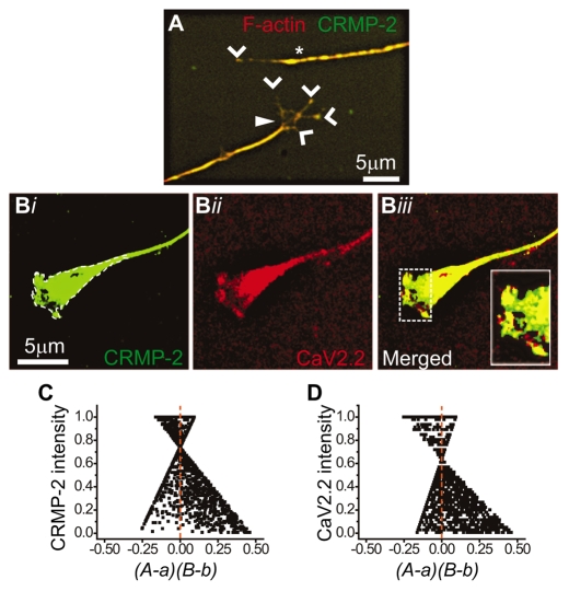
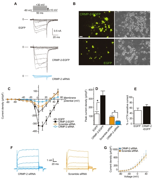
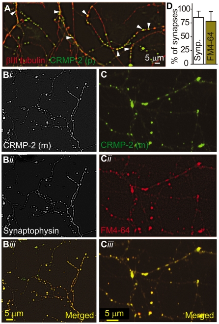
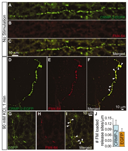
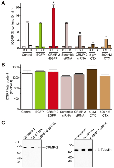
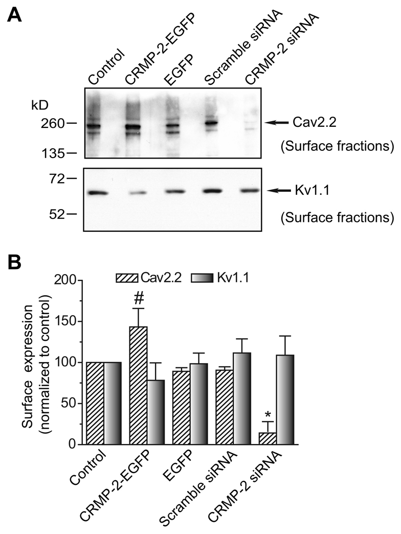
Collapsin response mediator proteins (CRMPs) mediate signal transduction of neurite outgrowth and axonal guidance during neuronal development. Voltage-gated Ca(2+) channels and interacting proteins are essential in neuronal signaling and synaptic transmission during this period. We recently identified the presynaptic N-type voltage-gated Ca(2+) channel (Cav2.2) as a CRMP-2-interacting partner. Here, we investigated the effects of a functional association of CRMP-2 with Cav2.2 in sensory neurons. Cav2.2 colocalized with CRMP-2 at immature synapses and growth cones, in mature synapses and in cell bodies of dorsal root ganglion (DRG) neurons. Co-immunoprecipitation experiments showed that CRMP-2 associates with Cav2.2 from DRG lysates. Overexpression of CRMP-2 fused to enhanced green fluorescent protein (EGFP) in DRG neurons, via nucleofection, resulted in a significant increase in Cav2.2 current density compared with cells expressing EGFP. CRMP-2 manipulation changed the surface levels of Cav2.2. Because CRMP-2 is localized to synaptophysin-positive puncta in dense DRG cultures, we tested whether this CRMP-2-mediated alteration of Ca(2+) currents culminated in changes in synaptic transmission. Following a brief high-K(+)-induced stimulation, these puncta became loaded with FM4-64 dye. In EGFP and neurons expressing CRMP-2-EGFP, similar densities of FM-loaded puncta were observed. Finally, CRMP-2 overexpression in DRG increased release of the immunoreactive neurotransmitter calcitonin gene-related peptide (iCGRP) by approximately 70%, whereas siRNA targeting CRMP-2 significantly reduced release of iCGRP by approximately 54% compared with control cultures. These findings support a novel role for CRMP-2 in the regulation of N-type Ca(2+) channels and in transmitter release.


Figures

Fig. 1.
Fig. 1.
CRMP-2 colocalizes with Cav2.2 in DRG growth cones. (A) Immunocytochemistry of DRG growth cones double-labeled with the growth-cone-enriched protein F-actin (red) and CRMP-2 (green). Note the extensive colocalization of both proteins in the lamellipodia of the collapsed (asterisk) and spread (closed arrowhead) growth cones. Staining of both proteins at the edges of the filopodia is also evident (open arrowheads). (B) Pseudocolor images of a DRG growth cone labeled with CRMP-2 (Bi) and Cav2.2 (Bii), with the merged image shown in (Biii). Inset: enlarged region of the peripheral zone of the growth cone showing extensive colocalization of CRMP-2 and Cav2.2. (C,D) The region demarcated by the white broken line in Bi generated scatter plots with strong right skews for CRMP-2 (C) and Cav2.2 (D). The ICQ value for the region shown was 0.11 (P=0<0.05).
Fig. 2.
Fig. 2.
CRMP-2 colocalizes with Cav2.2 in DRG neurons. (A,B) Immunocytochemistry with anti-CRMP-2 (A) and anti-Cav2.2 (B) antibodies in adult rat DRG neurons cultured for 7 DIV. (C) Merged pseudocolor image (CRMP-2, red; Cav2.2, green). (D,E) ICA of the soma surface (inset in C) generated scatter plots with strong right skews for both CRMP-2 (D) and Cav2.2 (E). The intensity correlation quotient (ICQ) values for the regions shown in the inset in C was 0.295 (P=0<0.05; juxtamembrane region of DRG soma). (F) Boxplot showing the ICQ values for CRMP-2:Cav2.2 staining from cell-surface soma (n=8). (G) V5-epitope-tagged CRMP-1 and CRMP-2 proteins were expressed in heterologous cells, immunoprecipitated with V5-tag antibody and immunoblotted with CRMP-2 (top blot) or V5 antibody (lower blot). The CRMP-2 antibody recognized a single band for CRMP-2 in immunoprecipitations from cells expressing CRMP-2 but not CRMP-1. (H,I) DRG lysates were immunoprecipitated (IP) with Cav2.2 and CRMP-2 antibodies and immunoblotted with Cav2.2 (H) and CRMP-2 antibodies (I). Input lane represents 5% of the starting material used for IP. Only the light chains (LC) of the IgG were observed with an anti-rabbit IgG (light-chain-specific) secondary antibody. A representative blot from four experiments is shown. Molecular weight markers are indicated in kDa.
Fig. 3.
Fig. 3.
CRMP-2 increases Ca2+ current density in DRG neurons. (A) Exemplar current traces obtained from DRG neurons transfected with EGFP, CRMP-2–EGFP or CRMP-2 siRNA. Currents were evoked by 50-millisecond steps in 10 mV increments applied from a holding potential of –80 mV (see voltage protocol above the traces). Current traces are shown for every 10-mV step between –80 and +30 mV. Bath solutions contained 1 μM TTX, 10 mM TEA and 1 μM nifedipine to block Na+, K+ and L-type voltage-gated Ca2+ channels, respectively. Lines labeled 0 indicate the zero-current level. (B) Representative fluorescence and differential interference contrast (DIC) images of CRMP-2–EGFP- and EGFP-transfected DRG neurons. Scale bar: 20 μm. (C) Current-voltage relationships for the currents shown in A as well as for currents from cells transfected with scramble siRNA. Peak currents were normalized to cell capacitance. The Ca2+ current density was significantly greater in CRMP-2-EGFP neurons than that in EGFP between 0 to +50 mV (P<0.05, Student's t-test). Values are mean ± s.e.m. and some error bars are smaller than the symbols. (D) Peak current density (pA/pF) measured at +10 mV for EGFP (n=6) is significantly smaller than that for neurons transfected with CRMP-2–EGFP (n=11) (**P<0.01, Student's t-test). Peak current density for CRMP-2 siRNA (n=6) is significantly smaller than for scramble siRNA (*P<0.05, Student's t-test). Peak current density values are not different between EGFP and scramble siRNA (P>0.2, Student's t-test). (E) Percent inhibition of Ca2+ current via Cav2.2, at 5 minutes post-perfusion with 1 μM ω-CTX, in DRGs expressing EGFP (n=4) and CRMP-2–EGFP (n=9). (F) Representative K+ currents from a DRG neuron transfected with CRMP-2 siRNA (left) and scramble siRNA (right) in response to 10-mV voltage steps from –80 to +60 mV from a holding potential of –60 mV. (G) Current-voltage relationship for DRG neurons treated with scramble and CRMP-2 siRNA (n=6 each). Peak K+ currents were not different between the two conditions for any of the voltages tested.
Fig. 4.
Fig. 4.
Effect of CRMP-2 on DRG Cav2.2 activation. (A) Normalized conductance (G) versus voltage for DRG neurons transfected with EGFP or CRMP-2–EGFP. Values are mean ± s.e.m. and most error bars are smaller than the symbols. (B) Plots of activation time constants (τactivation) were calculated from single exponential fits to the falling phase of the voltage-activated inward current. The mean activation time constants for CRMP-2–EGFP currents were significantly faster than those for EGFP (P<0.05, Student's t-test).
Fig. 5.
Fig. 5.
Effect of CRMP-2 on DRG Cav2.2 inactivation. (A) Top panel: voltage protocol. Currents were evoked by voltage steps from –80 to +40 mV in 10-mV increments prior to delivering a test pulse to +10 mV. Bottom two panels: exemplar current traces from EGFP and CRMP-2–EGFP transfected neurons. (B) Normalized test pulse peak current amplitude plotted against its preceding holding potential and fitted with the Boltzmann relation. (C) Plot of mean V50% for EGFP and CRMP-2–EGFP showed no difference between the groups.
Fig. 6.
Fig. 6.
Endogenous CRMP-2 colocalizes to synaptic boutons. (A) Representative confocal immunofluorescence image of DRGs stained with a monoclonal antibody against βIII tubulin (red) and polyclonal (p) CRMP-2 (green). CRMP-2 stains distinct punctate structures (arrowheads) along DRG neurites. (B) Confocal black and white image of DRG neurites double-labeled with a monoclonal CRMP-2 antibody (green) and a polyclonal synaptophysin antibody (red). The merged image shows that CRMP-2 protein localizes to synapses identified by the synaptophysin antibody. (C) DRGs grown for 7 DIV were loaded with FM4-64 (red) and then fixed and stained with a monoclonal (m) antibody against CRMP-2 and identified with an FITC-conjugated goat anti-mouse secondary antibody (green). Note the colocalization of CRMP-2 with FM4-64 in active synapses. (D) Quantification of the percentage of CRMP-2-positive puncta that were also labeled with synaptophysin (synp.) or FM4-64. A total of 468 (n=7 neurons) and 347 puncta (n=8 neurons) were counted for synaptophysin and FM4-64, respectively.
Fig. 7.
Fig. 7.
CRMP-2 does not affect synapse density in DRG neurons. (A-I) DRGs grown in culture for 5 DIV were transfected with CRMP-2–EGFP (A–F) or EGFP (G–I) and then incubated for 1 minute without (A–C) or with 90 mM KCl (D–I) for 1 minute to label release sites. Representative confocal images of neurites are shown. Note the lack of FM4-64 staining in the absence of stimulation. In high-K+-stimulated neurons, EGFP fluorescence of cells transfected with EGFP and CRMP-2–EGFP colocalizes with FM4-64 (I and F). (J) Quantification of the number of FM dye-loaded synapses in neurons transfected with EGFP or in cells transfected with CRMP-2–EGFP (CRMP-2). A total of 175 (n=9 EGFP neurons) and 184 puncta (n=13 CRMP-2–EGFP neurons) were counted.
Fig. 8.
Fig. 8.
CRMP-2 affects K+-stimulated transmitter release in DRG neurons. Adult mouse DRG neurons were maintained in culture for 5-7 days prior to the release experiments. (A) Bar graph of iCGRP release expressed as mean percent total iCGRP content of cells in each well ± s.e.m. (n=12-24 wells per condition). Neuropeptide release was measured from cells treated with normal HEPES buffer containing 3.5 mM KCl (basal, B), HEPES buffer containing 50 mM KCl (S), and again with HEPES buffer containing 3.5 mM KCl. The Cav2.2 inhibitor, ω-CTX, at 5.0 μM or 500 nM, was included in the 10 minutes prior to and throughout the high K+ exposures. Total inhibitor exposure time was 20 minutes. Asterisks (*) indicate statistically significant differences in iCGRP release between treatment groups and the control (no treatment) using an ANOVA with Dunnett's post-hoc test (P<0.05). The hash mark (#) indicates a statistically significant difference between the scramble- and CRMP-2-siRNA-treated cells (P<0.05; Student's t-test). In all cases, release stimulated by high extracellular K+ was significantly higher than basal release. (B) Total content of iCGRP measured at the end of the release experiment. There were no significant differences in iCGRP content between the conditions tested. (C) Western blot analysis with the indicated antibodies showing successful knockdown of CRMP-2 protein in neurons treated with 200 nM siRNAs for 4 days. Under the same conditions, expression of the neuronal protein β-tubulin remained unchanged. Lysates from untreated and scramble-siRNA-treated DRGs showed no differences in protein expression for either CRMP-2 or β-tubulin. Knockdown was assessed by quantifying densities of CRMP-2 for each treatment and then normalizing them to their respective β-tubulin densities (treatment CRMP-2 density/β-tubulin). This value was then divided by the normalized density obtained for the parallel untreated control (control CRMP-2 density/β-tubulin). For the blot shown, treatment with CRMP-2siRNA reduced CRMP-2 by ∼90% compared to treatment with scramble (Sc) siRNA. A representative blot from two experiments is shown.
Fig. 9.
Fig. 9.
CRMP-2 affects Cav2.2 surface expression. (A) DRG neurons grown in culture were transfected as indicated and then biotinylated with EZ-link Sulfo NHS-SS-biotin 2 days after transfection. Equal amounts of biotinylated proteins precipitated with streptavidin agarose beads were resolved by electrophoresis and subjected to immunoblot analysis with Cav2.2 (top blot) and Kv1.1 (bottom blot). (B) Amount of biotinylated (surface) Cav2.2 or Kv1.1 protein, calculated by densitometry from three experiments. Values were normalized to the untransfected (control) cells for each condition. The surface levels of Cav2.2 were significantly higher in CRMP-2-EGFP neurons than that in EGFP neurons (#P<0.05, Student's t-test). The surface Cav2.2 levels were significantly lower in neurons treated with CRMP-2 siRNA than in neurons treated with scramble siRNA (*P<0.05, Student's t-test). Levels of Kv1.1 were not significantly different between any of the conditions tested.

Similar articles

Cited by

References

    1. Alvarez, F. J., Kavookjian, A. M. and Light, A. R. (1993). Ultrastructural morphology, synaptic relationships, and CGRP immunoreactivity of physiologically identified C-fiber terminals in the monkey spinal cord. J. Comp Neurol. 329, 472-490. - PubMed
    1. Ambalavanar, R., Moritani, M., Moutanni, A., Gangula, P., Yallampalli, C. and Dessem, D. (2006). Deep tissue inflammation upregulates neuropeptides and evokes nociceptive behaviors which are modulated by a neuropeptide antagonist. Pain 120, 53-68. - PubMed
    1. Augustine, G. J. (2001). How does calcium trigger neurotransmitter release? Curr. Opin. Neurobiol. 11, 320-326. - PubMed
    1. Bahls, F. H., Lartius, R., Trudeau, L. E., Doyle, R. T., Fang, Y., Witcher, D., Campbell, K. and Haydon, P. G. (1998). Contact-dependent regulation of N-type calcium channel subunits during synaptogenesis. J. Neurobiol. 35, 198-208. - PubMed
    1. Behar, O., Mizuno, K., Badminton, M. and Woolf, C. J. (1999). Semaphorin 3A growth cone collapse requires a sequence homologous to tarantula hanatoxin. Proc. Natl. Acad. Sci. USA 96, 13501-13505. - PMC - PubMed

Publication types

Substances