. 2010 Jan 8;285(2):1343-57.
doi: 10.1074/jbc.M109.024703. Epub 2009 Nov 10.

Kidins220/ARMS modulates the activity of microtubule-regulating proteins and controls neuronal polarity and development

Affiliations

Kidins220/ARMS modulates the activity of microtubule-regulating proteins and controls neuronal polarity and development

Alonso M Higuero et al. J Biol Chem. .

Abstract

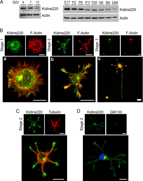
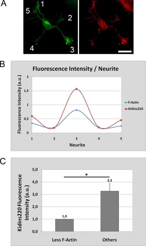
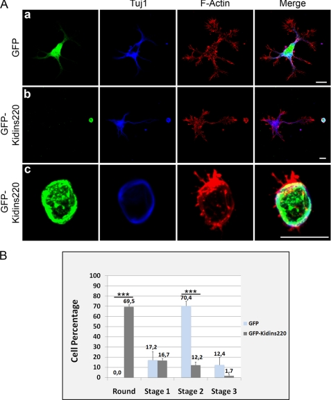
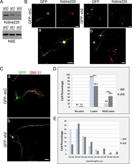
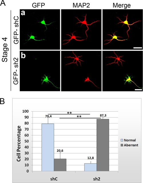
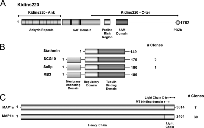
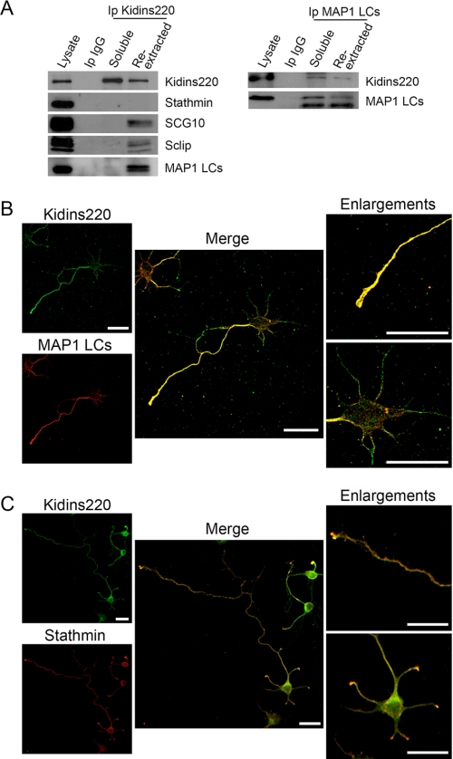
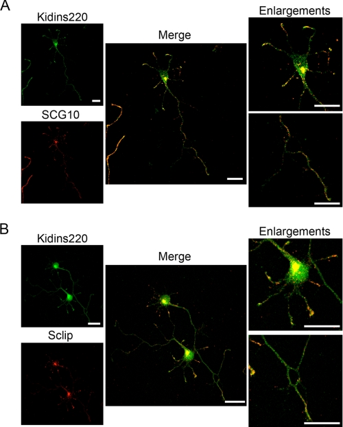
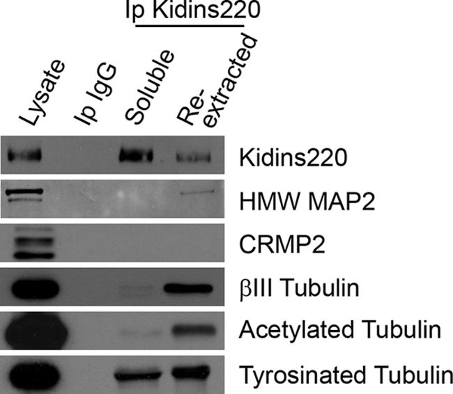
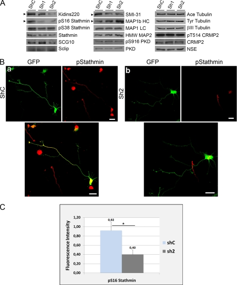
In order for neurons to perform their function, they must establish a highly polarized morphology characterized, in most of the cases, by a single axon and multiple dendrites. Herein we find that the evolutionarily conserved protein Kidins220 (kinase D-interacting substrate of 220-kDa), also known as ARMS (ankyrin repeat-rich membrane spanning), a downstream effector of protein kinase D and neurotrophin and ephrin receptors, regulates the establishment of neuronal polarity and development of dendrites. Kidins220/ARMS gain and loss of function experiments render severe phenotypic changes in the processes extended by hippocampal neurons in culture. Although Kidins220/ARMS early overexpression hinders neuronal development, its down-regulation by RNA interference results in the appearance of multiple longer axon-like extensions as well as aberrant dendritic arbors. We also find that Kidins220/ARMS interacts with tubulin and microtubule-regulating molecules whose role in neuronal morphogenesis is well established (microtubule-associated proteins 1b, 1a, and 2 and two members of the stathmin family). Importantly, neurons where Kidins220/ARMS has been knocked down register changes in the phosphorylation activity of MAP1b and stathmins. Altogether, our results indicate that Kidins220/ARMS is a key modulator of the activity of microtubule-regulating proteins known to actively regulate neuronal morphogenesis and suggest a mechanism by which it contributes to control neuronal development.


Figures

FIGURE 1.
FIGURE 1.
Kidins220/ARMS expression and distribution in hippocampal neurons in vitro. A, lysates from different developmental stages (DIV) of rat hippocampal neurons in culture or rat brain from embryonic (E), postnatal (P, days), or adult (M; months) animals were immunoblotted with anti-Kidins220/ARMS antibody. β-Actin was used as a control. B, hippocampal neurons were fixed at stage 1 (a), 2 (b), or 3 (c) and immunostained with phalloidin-TRITC (F-Actin, red). Scale bar, 10 μm. C and D, hippocampal neurons were fixed at stage 2 and immunostained with anti-Kidins220/ARMS (green) and α-tubulin (C; red) or the Golgi marker GM130 (D; red). Note that Kidins220/ARMS co-localizes with the Golgi apparatus in the juxtanuclear region. Nuclei stained with DAPI (blue) staining are shown in the merged image. Scale bar, 10 μm.
FIGURE 2.
FIGURE 2.
Correlation between Kidins220/ARMS and F-actin levels in stage 2 neurons. A, stage 2 hippocampal neurons were fixed and immunostained with anti-Kidins220/ARMS (green) and phalloidin-Texas Red (F-Actin, red). Scale bar, 20 μm. B, quantification of the relative fluorescence intensity of Kidins220/ARMS and F-actin for each numbered neurite of a representative stage 2 hippocampal neuron (shown in A). Note the high degree of correlation between the intensities of both signals. C, in a stage 2 hippocampal neuron population, the single neurite tip with lowest actin filament content had the lowest Kidins220/ARMS concentration, and those neurites with more F-actin had a 3.3-fold higher intensity for Kidins220/ARMS signal (n = 40, three independent experiments). Statistical significance was evaluated by the Student's unpaired t test (*, p < 0.05). a.u., arbitrary units.
FIGURE 3.
FIGURE 3.
Ectopic expression of GFP-Kidins220/ARMS hampers early neuronal development. A, hippocampal neurons transfected after dissection with GFP as control (a) or GFP-Kidins220/ARMS (b and c) were fixed at 1.5 DIV and immunostained for the neuronal marker βIII-tubulin/Tuj1 (blue) and phalloidin-TRITC (F-Actin, red). Scale bar, 10 μm. B, quantification of the number of rounded, stage 1, 2, or 3 neurons after Kidins220/ARMS overexpression relative to control GFP-transfected cells. The data shown are the means ± S.E. of three independent experiments (GFP n = 184 cells, GFP-Kidins220/ARMS n = 170 cells), and statistical significance was evaluated by Student's unpaired t test (***, p < 0.001).
FIGURE 4.
FIGURE 4.
Down-regulation of Kidins220/ARMS in hippocampal neurons induces the formation of multiple axon-like processes. A, PC12 cells were transfected with control shRNA (GFP-shC) or Kidins220/ARMS-specific shRNA (GFP-sh1 and -sh2) vectors. After 96 h, cells were lysed, and Kidin220/ARMS knockdown was examined by immunoblot analysis. Neuronal specific enolase (NSE) levels were used as control. B, hippocampal neurons were transfected with GFP-shC (a) or GFP-sh2 (b) and immunostained for Kidins220/ARMS (red). GFP-sh2 neurons showed decreased Kidins220/ARMS labeling. Merged images are also shown (a and b). Scale bar, 20 μm. C, hippocampal neurons transfected with GFP-shC (a) or GFP-sh2 (b) and fixed at stage 3 were immunostained with the axonal marker SMI-31 (red). Only merged images are shown. Early down-regulation of Kidins220/ARMS increased the number of SMI-31-positive processes per neuron. Scale bar, 20 μm. D, quantification of the percentage of hippocampal neurons bearing several axon-like processes after knocking down Kidins220/ARMS (sh2) compared with control (shC). The data shown are the means ± S.E. of three independent experiments (shC n = 148 cells, sh2 n = 128 cells), and statistical significance was evaluated by Student's unpaired t test (**, p < 0.01). E, frequency histogram of the percentage of hippocampal neurons with distinct axonal lengths in control or in Kidins220/ARMS knockdown cells (shC n = 100 cells, sh2 n = 100 cells).
FIGURE 5.
FIGURE 5.
Down-regulation of Kidins220/ARMS in hippocampal neurons alters normal dendrite development. A, hippocampal neurons were transfected with GFP-shC (a) as control or GFP-sh2 Kidins220/ARMS-specific shRNA (b). After 5 DIV, neurons were fixed and immunostained for the dendritic marker MAP2 (red). Similar to non-transfected neurons, GFP-shC neurons developed normal dendrites, whereas GFP-sh2 neurons displayed aberrant MAP2-positive processes. Compare the green neuron where Kidins220/ARMS has been knocked down with the neighbor neuron. Scale bar, 20 μm. B, quantification of the percentage of neurons at stage 4 bearing normal or aberrant dendritic processes. The data shown are the means ± S.E. of three independent experiments (shC n = 100 cells, sh2 n = 100 cells), and statistical significance was evaluated by Student's unpaired t test (**, p < 0.01).
FIGURE 6.
FIGURE 6.
Kidins220/ARMS domains used as baits in yeast two-hybrid assays and the interacting proteins identified. A, the 11 ankyrin repeats present at the Kidins220/ARMS N-terminal region (Kidins220-Ank) as well as its C-terminal region containing a proline-rich region, a SAM domain, and a PDZ binding motif (Kidins220-C-ter) were used as baits to screen a mouse brain cDNA library. B, schematic representation of the domain structure for the stathmin family members identified as Kidins220/ARMS-interacting proteins. All members contain the highly conserved regulatory and tubulin binding domains. SCG10, Sclip, and RB3 also possess an N-terminal membrane-anchoring domain absent in stathmin. Three independent clones of SCG10 and one clone of Sclip were identified. C, schematic representation of the domain structure for MAP1a and MAP1b. These proteins consist of a dimer between a heavy chain and a light chain derived from the same polypeptide. A very high number of clones of MAP1 were identified as Kinds220/ARMS-interacting proteins (37 in total).
FIGURE 7.
FIGURE 7.
Kidins220/ARMS interacts with SCG10, Sclip, and MAP1 in neurons. Cortical neurons were lysed to obtain a soluble fraction, and the insoluble pellet was re-extracted in order to immunoprecipitate Kidins220/ARMS or MAP1 LCs from both fractions (soluble and re-extracted). A, Kidins220/ARMS immunocomplexes were analyzed for the presence of Kidins220/ARMS (positive control) and stathmin, SCG10, Sclip, and MAP1 LCs by immunoblot. As a negative control, soluble lysates were also immunoprecipitated with an antibody against an IgG (Ip IgG). MAP1 LC immunocomplexes were analyzed for the presence of Kidins220/ARMS and MAP1 LCs (positive control) by immunoblot. As a negative control, soluble lysates were also immunoprecipitated with an antibody against an IgG. B, hippocampal neurons fixed at stage 3 were immunostained for Kidins220/ARMS (green) and MAP1 LCs (red), and their colocalization was analyzed by confocal microscopy. Both proteins colocalize predominantly in the axon and its growth cone (see enlargements). C, stage 3 hippocampal neurons were immunostained with anti-Kidins220/ARMS (green) and anti-stathmin (red) antibodies. The inserts show how stathmin and Kidins220/ARMS colocalize in the axonal and dendritic growth cones. A single 0.7-μm section for each channel and their merged image is shown. Scale bar, 20 μm.
FIGURE 8.
FIGURE 8.
Kidins220/ARMS colocalizes with SCG10 and Sclip in hippocampal neurons. A and B, hippocampal neurons fixed at stage 3 were immunostained for Kidins220/ARMS (green) and SCG10 or Sclip (red), and their colocalization was analyzed by confocal microscopy. The enlargements show how these two stathmin family members and Kidins220/ARMS colocalize predominantly in the dendritic growth cones and in a juxtanuclear region. A single 0.7-μm section for each channel and their merged image is shown. Scale bar, 20 μm.
FIGURE 9.
FIGURE 9.
Kidins220/ARMS interacts with MAP2 and tubulin in cortical neurons. Soluble and re-extracted fractions form cortical neurons were used to immunoprecipitate Kidins220/ARMS. As a negative control, similar soluble lysates were incubated with an antibody against a non-relevant IgG chain (Ip IgG). The different immunocomplexes were analyzed for the capacity of Kidins220/ARMS to immunoprecipitate MAP2 and CRMP2 as well as βIII-tubulin or the post-translationally modified (acetylated or tyrosinated) α-tubulin. As a positive control, Kidins220/ARMS is present in its own immunocomplexes.
FIGURE 10.
FIGURE 10.
Kidins220/ARMS knockdown modulates the phosphorylation of stathmins and MAP1b-HC. A, hippocampal neurons were transfected with control (shC) or Kidins220/ARMS-specific (sh1 and sh2) shRNA vectors, and Kidins220/ARMS protein levels were determined after 4 DIV by immunoblot analysis. The same lysates were probed for phosphorylated forms of stathmin (Ser(P)-16 (pS16) and Ser(P)-38 (pS38)); total stathmin, SCG10, and Sclip; the phosphorylated form of MAP1b (SMI-31 antibody, 320–340 kDa band); total MAP1b HC and LCs; MAP2; Ser(P)-916 (pS916) PKD; and total PKD as well as for the acetylated and tyrosinated forms of α-tubulin, βIII tubulin, Thr(P)-514 (pT514) CRMP2, and total CRMP2. Neuronal specific enolase (NSE) is shown as a control. The arrowheads indicate immunoblots for which significant signal intensity changes were observed. One representative immunoblot of three independent experiments is shown. B, hippocampal neurons were transfected with GFP-shC (a) as control or GFP-sh2 Kidins220/ARMS-specific shRNA (b) and immunostained with antibodies against Ser(P)-16-stathmin (red). GFP signal served to identify neurons transfected with the different shRNAs. Scale bar, 20 μm. C, quantification of the relative fluorescence intensity for Ser(P)-16-stathmin on GFP-positive (transfected) neurons with respect to GFP-negative (untransfected) cells. Although shC-transfected neurons display a nearly identical intensity of Ser(P)-16-stathmin as untransfected neurons (ratio is almost 1), Ser(P)-16-stathmin decreases significantly in sh2-transfected neurons. The data shown are the means ± S.E. of three independent experiments (shC n = 52 cells, sh2 n = 74 cells), and statistical significance was evaluated by Student's unpaired t test (*, p < 0.05).

Similar articles

Cited by

References

    1. Arimura N., Kaibuchi K. (2007) Nat. Rev. Neurosci. 8, 194–205 - PubMed
    1. da Silva J. S., Dotti C. G. (2002) Nat. Rev. Neurosci. 3, 694–704 - PubMed
    1. Da Silva J. S., Hasegawa T., Miyagi T., Dotti C. G., Abad-Rodriguez J. (2005) Nat. Neurosci. 8, 606–615 - PubMed
    1. Horton A. C., Ehlers M. D. (2003) Neuron 40, 277–295 - PubMed
    1. Bradke F., Dotti C. G. (2000) Curr. Opin. Neurobiol. 10, 574–581 - PubMed

Publication types

MeSH terms

LinkOut - more resources