. 2010 Jan 1;285(1):216-25.
doi: 10.1074/jbc.M109.041640. Epub 2009 Nov 10.

NFkappaB p50-CCAAT/enhancer-binding protein beta (C/EBPbeta)-mediated transcriptional repression of microRNA let-7i following microbial infection

Affiliations

NFkappaB p50-CCAAT/enhancer-binding protein beta (C/EBPbeta)-mediated transcriptional repression of microRNA let-7i following microbial infection

Steven P O'Hara et al. J Biol Chem. .

Abstract

MicroRNAs, central players of numerous cellular processes, regulate mRNA stability or translational efficiency. Although these molecular events are established, the mechanisms regulating microRNA function and expression remain largely unknown. The microRNA let-7i regulates Toll-like receptor 4 expression. Here, we identify a novel transcriptional mechanism induced by the protozoan parasite Cryptosporidium parvum and Gram(-) bacteria-derived lipopolysaccharide (LPS) mediating let-7i promoter silencing in human biliary epithelial cells (cholangiocytes). Using cultured cholangiocytes, we show that microbial stimulus decreased let-7i expression, and promoter activity. Analysis of the mechanism revealed that microbial infection promotes the formation of a NFkappaB p50-C/EBPbeta silencer complex in the regulatory sequence. Chromatin immunoprecipitation assays (ChIP) demonstrated that the repressor complex binds to the let-7i promoter following microbial stimulus and promotes histone-H3 deacetylation. Our results provide a novel mechanism of transcriptional regulation of cholangiocyte let-7i expression following microbial insult, a process with potential implications for epithelial innate immune responses in general.


Figures
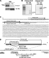
FIGURE 1.
FIGURE 1.
Identification of the primary let-7i transcript. A, Northern blot for the primary let-7i transcript identified an approximate 760-nucleotide RNA in uninfected H69 cells, whereas C. parvum infection diminishes the expression of this RNA. 18 S rRNA was used as a loading control. B, RACE-PCR, using polyadenylated enriched RNA, was used to identify the primary let-7i transcript. A 100-bp let-7i specific amplicon was identified using 5′-RACE and sequence verified. A single intense band was observed at ∼750 bp in our 3′-nested RACE PCR. This let-7i specific band was sequence verified. C, primary let-7i sequence was identified in cholangiocytes by RACE-PCR with subsequent cloning and sequencing. The 5′ and 3′ gene-specific primers (GSPs) we generated based on the let-7i precursor sequence (bold). The dashed box indicates the mature let-7i sequence, and the predicted polyadenylation signal is underlined. D, let-7i primary transcript is located on chromosome 12 and exists within an intron of an EST (DA092355) identified in human cerebellum. Let-7i is flanked upstream by an open reading frame, which lies in the opposite orientation. MON2, which encodes a large guanine nucleotide exchange factor, terminates ∼6,000 base pairs upstream whereas the protein phosphatase PPM1H lies in the opposite orientation and terminates ∼40,000 base pairs downstream.
FIGURE 2.
FIGURE 2.
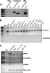
Expression analyses of genes located on chromosome 12q14. A, rtPCR was used to detect the EST, DA092355, from cerebellum. This transcript was not expressed in H69 cells. 18 S rRNA was amplified as control. B, primary let-7i transcript was detected by rtPCR in all gastrointestinal tissues assessed, whereas the EST DA092355 was not detected in any of these tissues. C, genes flanking let-7i on chromosome 12 including MON2, C12ORF61, and PPM1H, were identified in cerebellum by rtPCR, yet were not detected in basal or microbe-stimulated H69 cells.
FIGURE 3.
FIGURE 3.
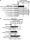
Functional analyses of the let-7i Dual-Luciferase reporter assay. A, 2,461 base pairs upstream of the identified let-7i transcription start site were cloned into the luciferase expression plasmid pGL4.22. This putative let-7i promoter drives transcription over 10-fold compared with empty vector control. Serial truncations (Δ1-Δ5) identify a region between −1110 and −961 that is necessary for basal transcription. Two putative NFκB binding sites (−619 and −657) and a C/EBPβ binding site (−1166) were identified within the cloned promoter fragment. B, let-7i promoter-driven luciferase construct is responsive to microbial stimulus. C. parvum infection or LPS treatment reduces luciferase expression over 50% compared with uninfected cells. The observed microbial-induced suppression is diminished following pretreatment of the cells with the NFκB inhibitors MG132 and SN50. Expression was calculated as the relative let-7i-driven Firefly luciferase to transfection control Renilla luciferase and presented as fold-change compared with empty vector pGL4.22 Firefly luciferase to Renilla luciferase, which was arbitrarily set to one. Data are represented as mean ± S.E. from three separate experiments with at least n = 4 for each experiment. *, p < 0.05 compared with control luciferase by ANOVA.
FIGURE 4.
FIGURE 4.
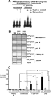
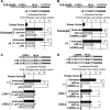
Immune-associated transcription factors interact with the putative let-7i promoter following microbial stimulus. A, electrophoretic mobility shift assays were used to demonstrate interactions between nuclear factors and the let-7i promoter following infection. An oligonucleotide containing the proximal NFκB binding site did not shift when the nuclear extract from basal, uninfected cells was used. However, the same oligonucleotide demonstrated less mobility through the gel when the nuclear extract from C. parvum-infected cells was used. This interaction could be competed away using unlabeled oligonucleotide competimer. B, ChIP analysis demonstrated that both NFκB p50 (but not p65) and C/EBPβ interact with the let-7i promoter following microbial stimulus. ChIP analyses for the interleukin-8 promoter were performed as a positive control for p65 with both uninfected and C. parvum-infected H69 cells. C, quantitative PCR ChIP was also performed to verify the increased interaction between the let-7i promoter and the transcription factors NFκB p50 and C/EBPβ. A standard curve was generated using amplicons of a known copy number. The data are presented as the relative amount of the immunoprecipitated fraction compared with total input DNA. Data are represented as mean ± S.E. from four separate experiments. *, p < 0.05 compared with respective uninfected controls by ANOVA.
FIGURE 5.
FIGURE 5.
Manipulation of NFκB p50 and C/EBPβ expression reciprocally affects reporter gene expression. A, the 2,461-bp let-7i promoter increased transcription over 12-fold compared with the pGL4.22 empty vector. Overexpression (oe) of p50 significantly decreased luciferase expression over 25%, whereas C/EBPβ overexpression diminished luciferase expression over 75%. Concurrent overexpression of these transcription factors synergistically represses luciferase expression from this promoter. B, same plasmid construct was used to demonstrate that RNA-mediated silencing of these transcription factors reciprocally affected luciferase expression driven from this promoter. Furthermore, siRNA-induced repression of p50 or C/EBPβ inhibited microbial-induced reduction of luciferase driven by the let-7i promoter. Data are represented as mean ± S.E. from three separate experiments with at least n = 4 for each experiment. *, p < 0.05 between comparisons indicated, using ANOVA.
FIGURE 6.
FIGURE 6.
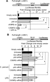
The consensus κB binding sites within the let-7i promoter confer responsiveness to microbial stimulus. A, the 2,461 bp promoter and a truncation of this promoter (Δ4), which lacks the predicted C/EBPβ binding site, was used to assess the functional significance of this portion of the cloned promoter. Both the full-length promoter (2,461 bp) and the truncated promoter were responsive to microbial stimulus. C. parvum infection or LPS treatment reduced luciferase expression ∼50% when either promoter construct was used. B, similarly, both the full-length promoter and the truncated promoter were responsive to p50 or C/EBPβ overexpression. Overexpression of p50 reduced luciferase expression ∼50% when either promoter construct was used, whereas C/EBPβ overexpression resulted in a greater than 5-fold reduction in luciferase expression when either promoter construct was used. C, Δ1 construct, which contains both the C/EBPβ and NFκB sites, was used to assess the functional significance of the putative κB binding sites. The Δ1 construct was responsive to microbial stimulus whereas the promoter constructs harboring a deletion of either predicted κB binding site (Δ1M-1 or Δ1M-2) abrogated the microbe-induced suppression of luciferase expression. D, Δ1 construct was also utilized to assess the functional significance of the κB binding sites with respect to NFκB p50 and C/EBPβ overexpression. C/EBPβ overexpression resulted in diminished luciferase expression in each of the promoter constructs. The Δ1 construct was responsive to p50 overexpression. Conversely, p50 overexpression does not significantly affect luciferase expression when the distal or proximal NFκB binding sites are eliminated, suggesting that these sites confer responsiveness to p50 overexpression. Expression was calculated as the relative let-7i-driven firefly luciferase to transfection control Renilla luciferase and presented as fold-change compared with empty vector pGL4.22 firefly luciferase to Renilla luciferase, which was arbitrarily set to one. Data are represented as mean ± S.E. from four separate experiments with at least n = 4 for each experiment. *, p < 0.05 compared with respective untreated controls by ANOVA.
FIGURE 7.
FIGURE 7.
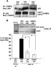
NFκB p50 and C/EBPβ interact and overexpression of either induces let-7i promoter deacetylation. A, immunoprecipitations demonstrate the interactions between NFκB p50 and C/EBPβ. Both the immunoprecipitation of C/EBPβ with subsequent blot for p50 and the converse immunoprecipitation of p50 and blot for C/EBPβ demonstrate that these proteins physically interact in cholangiocytes. Both the Lap1 and Lap2 isoforms were detected in cholangiocytes. B, ChIP analyses for acetylated Histone H3 were performed in both control, uninfected, and in cholangiocytes overexpressing either NFκB p50 or C/EBPβ. Overexpression of either transcription factor significantly (p < 0.01) decreased the detection of acetylated Histone H3 at the let-7i promoter. The data are presented as the relative amount of the immunoprecipitated fraction compared with total input DNA. Data are represented as mean ± S.E. from three separate experiments. *, p < 0.01 compared with H69 control by ANOVA.

Similar articles

Cited by

References

    1. Harada K., Ohba K., Ozaki S., Isse K., Hirayama T., Wada A., Nakanuma Y. (2004) Hepatology 40, 925–932 - PubMed
    1. Chen X. M., O'Hara S. P., LaRusso N. F. (2008) Immunol. Cell Biol. 86, 497–505 - PMC - PubMed
    1. Chen X. M., Splinter P. L., O'Hara S. P., LaRusso N. F. (2007) J. Biol. Chem. 282, 28929–28938 - PMC - PubMed
    1. Yokoyama T., Komori A., Nakamura M., Takii Y., Kamihira T., Shimoda S., Mori T., Fujiwara S., Koyabu M., Taniguchi K., Fujioka H., Migita K., Yatsuhashi H., Ishibashi H. (2006) Liver Int. 26, 467–476 - PubMed
    1. Chen X. M., Levine S. A., Splinter P. L., Tietz P. S., Ganong A. L., Jobin C., Gores G. J., Paya C. V., LaRusso N. F. (2001) Gastroenterology 120, 1774–1783 - PubMed

Publication types

MeSH terms