. 2010 Jan 15;285(3):1980-8.
doi: 10.1074/jbc.M109.016741. Epub 2009 Nov 10.

Farnesyl pyrophosphate inhibits epithelialization and wound healing through the glucocorticoid receptor

Affiliations

Farnesyl pyrophosphate inhibits epithelialization and wound healing through the glucocorticoid receptor

Sasa Vukelic et al. J Biol Chem. .

Abstract

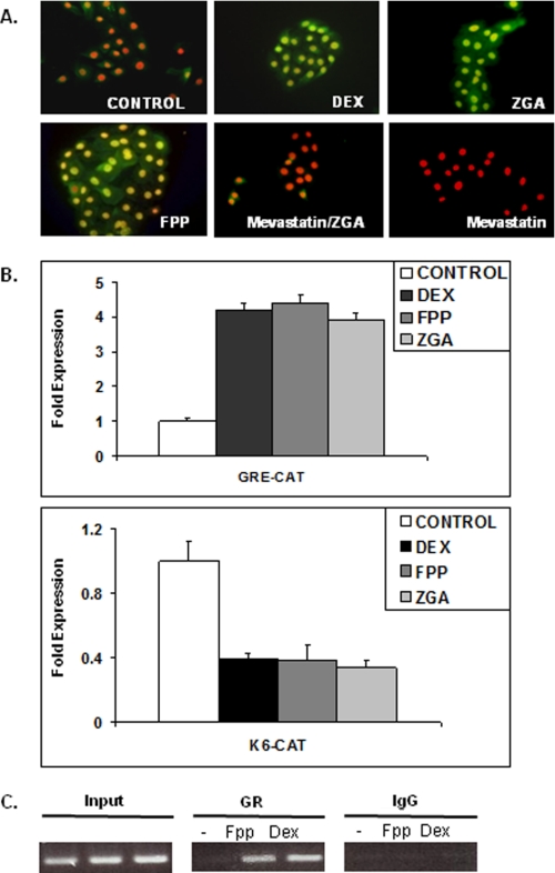
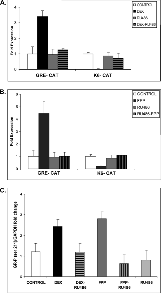
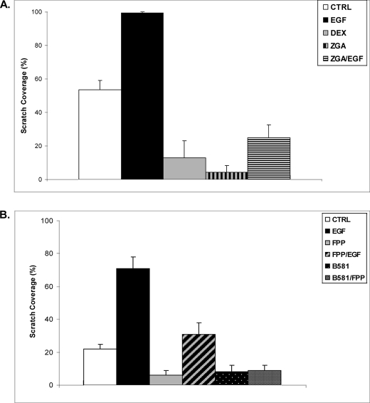
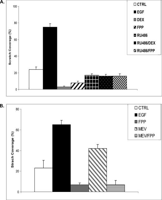
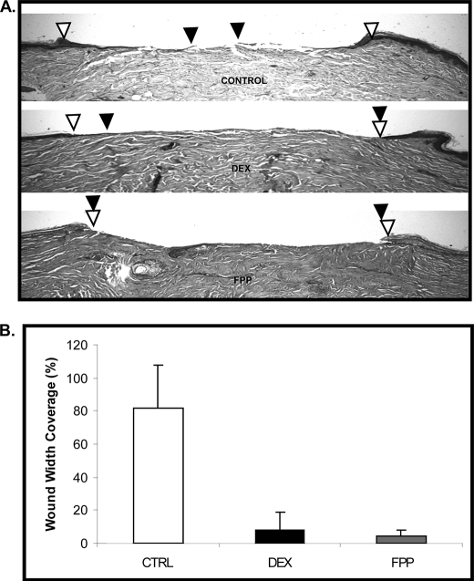
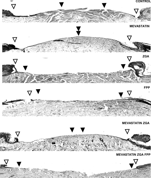
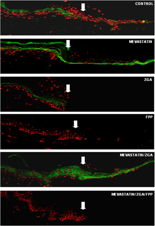
Farnesyl pyrophosphate (FPP), a key intermediate in the mevalonate pathway and protein farnesylation, can act as an agonist for several nuclear hormone receptors. Here we show a novel mechanism by which FPP inhibits wound healing acting as an agonist for glucocorticoid receptor (GR). Elevation of endogenous FPP by the squalene synthetase inhibitor zaragozic acid A (ZGA) or addition of FPP to the cell culture medium results in activation and nuclear translocation of the GR, a known wound healing inhibitor. We used functional studies to evaluate the effects of FPP on wound healing. Both FPP and ZGA inhibited keratinocyte migration and epithelialization in vitro and ex vivo. These effects were independent of farnesylation and indicate that modulation of FPP levels in skin may be beneficial for wound healing. FPP inhibition of keratinocyte migration and wound healing proceeds, in part, by repression of the keratin 6 gene. Furthermore, we show that the 3-hydroxy-3-methylglutaryl-CoA-reductase inhibitor mevastatin, which blocks FPP formation, not only promotes epithelialization in acute wounds but also reverses the effect of ZGA on activation of the GR and inhibition of epithelialization. We conclude that FPP inhibits wound healing by acting as a GR agonist. Of special interest is that FPP is naturally present in cells prior to glucocorticoid synthesis and that FPP levels can be further altered by the statins. Therefore, our findings may provide a better understanding of the pleiotropic effects of statins as well as molecular mechanisms by which they may accelerate wound healing.


Figures

FIGURE 1.
FIGURE 1.
FPP mediates activation and function of GR in human keratinocytes. A, FPP activates GR. Primary human keratinocytes stained with anti-Ser(P)211 GR antibody reveal localization and activation of GR. This antibody recognizes ligand-induced phosphorylation at Ser211. The nuclei were visualized by propidium iodide staining. Weak Ser(P)211 GR immunoreactivity was observed in the cytoplasm and nuclei of untreated cells. In contrast, strong signal was evident in the nuclei of all treated cells: DEX, FPP, and ZGA, which elevates endogenous FPP levels. Mevastatin abolishes ZGA-induced activation of GR by preventing accumulation of endogenous FPP. B, FPP activates GR-mediated transcriptional regulation in keratinocytes. Transfection experiments of primary human keratinocytes with the K6-CAT and GRE-CAT reporters are shown. The data are presented as relative CAT activity, a measure of actual CAT activity normalized for total protein. The results show that DEX, ZGA, or FPP treatment lead to repression of the K6 promoter. Similar to DEX, FPP or ZGA also stimulated the GRE-CAT reporter. C, FPP targets the K6 promoter through GR. HEK were either untreated (−) or treated with 1 μm DEX or 10 μm FPP. Similar amounts of genomic DNA (Input) was used in each treatment. K6 promoter sequences were amplified by PCR after immunoprecipitation with anti-GR antibody or rabbit IgG.
FIGURE 2.
FIGURE 2.
FPP-activated GR is transcriptionally active. A and B, transfection experiments of HEK with the K6-CAT and GRE-CAT reporters are shown. The data are presented as relative CAT activity, a measure of actual CAT activity normalized to total protein. The results show that DEX and FPP treatment lead to repression of the K6 promoter and stimulation of the GRE-CAT reporter. The effect of both DEX and FPP was reversed with RU486 antagonist of GR. C, quantitative analysis of immunoblotting with anti-Ser(P)211 GR antibody normalized to GAPDH and plotted as fold change in GR phosphorylation. HEK were incubated in the presence or absence of 1 μm DEX, 10 μm FPP, 10 μm RU486, or a combination for 4 h. Both DEX and FPP induce an increase in Ser211 GR phosphorylation, which was reversed with RU486.
FIGURE 3.
FIGURE 3.
FPP blocks keratinocyte migration. A, ZGA and DEX inhibit keratinocyte migration in the wound scratch assay. EGF treatment stimulated keratinocyte migration, and after 48 h cells covered the scratch completely. When present together, ZGA blocks EGF-stimulated keratinocyte migration. The average coverage of the scratch widths in percentage is presented by the histograms. B, HEK were wounded by scratch and incubated with or without, EGF, FPP, EGF + FPP, B581, or B581 + FPP. Keratinocyte migration was monitored for 48 h. FPP shows the same inhibitory effect on keratinocyte migration as ZGA. Interestingly, treatment either with B581 alone or with FPP had the same effect, indicating that enhanced protein farnesylation mediated through increased FPP in cells does not inhibit migration. CTRL, control.
FIGURE 4.
FIGURE 4.
FPP inhibits HEK migration, whereas mevastatin promotes it. A, DEX (1 μm) and FPP (10 μm) inhibit keratinocyte migration in the wound scratch assay. When present together, RU486 (10 μm) reverses both DEX and FPP inhibition of keratinocyte migration. B, mevastatin (MEV, 10 μm) increases keratinocyte migration in the wound scratch assay. Simultaneous treatment with FPP (10 μm) abolishes the effect of mevastatin and leads to a similar inhibition of migration as a FPP treatment alone. EGF treatment served as a positive control (CTRL) in both experiments. As expected, it stimulated keratinocyte migration. The average coverage of the scratch widths in percentage is presented by the histograms.
FIGURE 5.
FIGURE 5.
FPP inhibits epidermal wound healing. A, FPP completely inhibited epithelialization in a human skin organ culture model. Topical DEX treatment delayed epithelialization when compared with control (CTRL) untreated skin. The open arrowheads indicate wound edges after initial wounding, whereas solid arrowheads point to the epithelialized edges of the migrating fronts 4 days after the wounding. B, the experiment shown in Fig. 5 was repeated three times in triplicate using human skin obtained from three different donors. Quantification of wound epithelialization using planimetry is shown.
FIGURE 6.
FIGURE 6.
FPP inhibits epithelialization in skin organ culture, whereas mevastatin reverses this inhibition and promotes wound closure. Open arrowheads indicate wound edges after initial wounding, whereas solid arrowheads point at the epithelialized edges of the migrating fronts 4 days after wounding. Mevastatin improved epithelialization by decreasing endogenous levels of FPP. Both FPP and ZGA treatment completely inhibited epithelialization when compared with control untreated skin, whereas mevastatin reversed the inhibitory effect of ZGA.
FIGURE 7.
FIGURE 7.
Altering endogenous levels of FPP modulates epithelialization through inhibition of K6, whereas mevastatin reverses it. Shown is immunolocalization of K6 (green) at the wound edge and epithelial tongue after injury to epidermis in the human skin organ culture model. The white arrows indicate the initial wound edges. Mevastatin induces the expression of K6 at the wound edge. Both ZGA and FPP reduce the levels of K6 at the wound margin and inhibit epithelialization. Mevastatin reverses the effect of ZGA because of an upstream block in mevalonate pathway. The addition of exogenous FPP restores inhibition of wound healing and K6 expression.

References

    1. Brem H., Tomic-Canic M. (2007) J. Clin. Invest. 117, 1219–1222 - PMC - PubMed
    1. Pastar I., Stojadinovic O., Tomic-Canic M. (2008) Surg. Technol. Int. 17, 105–112 - PubMed
    1. Tomic-Canic M., Magnus S. A., Oscar M. A. (2004) in The Epidermis in Wound Healing (Rovee D. T., Maibach H. I. eds) pp. 25–57, CRC Press LLC, Boca Raton, FL
    1. Tomic-Canic M., Komine M., Freedberg I. M., Blumenberg M. (1998) J. Dermatol. Sci. 17, 167–181 - PubMed
    1. Freedberg I. M., Tomic-Canic M., Komine M., Blumenberg M. (2001) J. Invest. Dermatol. 116, 633–640 - PubMed

Publication types

MeSH terms

LinkOut - more resources