. 2009 Nov;21(11):3623-40.
doi: 10.1105/tpc.109.068791. Epub 2009 Nov 10.

Efficient operation of NAD(P)H dehydrogenase requires supercomplex formation with photosystem I via minor LHCI in Arabidopsis

Affiliations

Efficient operation of NAD(P)H dehydrogenase requires supercomplex formation with photosystem I via minor LHCI in Arabidopsis

Lianwei Peng et al. Plant Cell. 2009 Nov.

Abstract

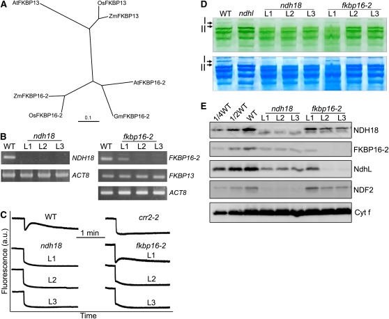
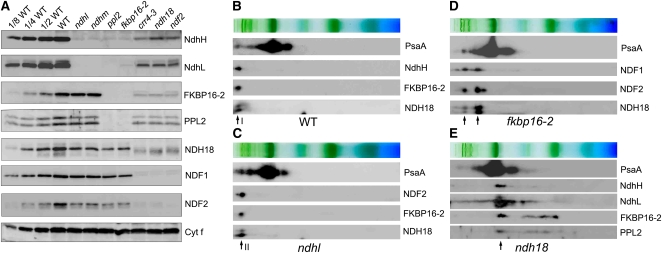
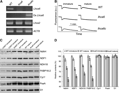
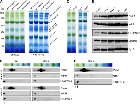
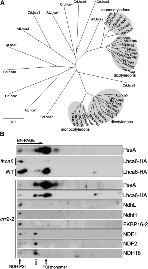
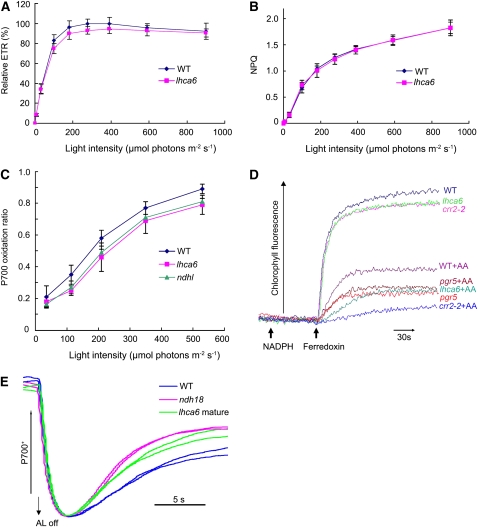
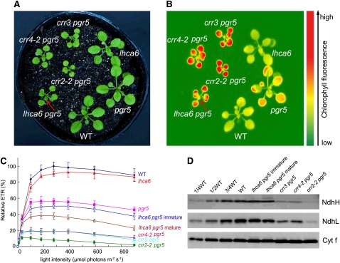
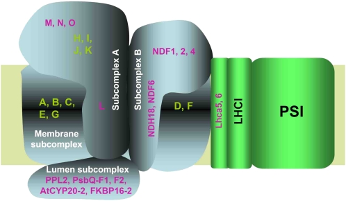
In higher plants, the chloroplast NAD(P)H dehydrogenase (NDH) complex mediates photosystem I (PSI) cyclic and chlororespiratory electron transport. We reported previously that NDH interacts with the PSI complex to form a supercomplex (NDH-PSI). In this study, NDH18 and FKBP16-2 (FK506 Binding Protein 16-2), detected in the NDH-PSI supercomplex by mass spectrometry, were shown to be NDH subunits by the analysis of their knockdown lines. On the basis of extensive mutant characterization, we propose a structural model for chloroplast NDH, whereby NDH is divided into four subcomplexes. The subcomplex A and membrane subcomplex are conserved in cyanobacteria, but the subcomplex B and lumen subcomplex are specific to chloroplasts. Two minor light-harvesting complex I proteins, Lhca5 and Lhca6, were required for the full-size NDH-PSI supercomplex formation. Similar to crr pgr5 double mutants that completely lack cyclic electron flow activity around PSI, the lhca6 pgr5 double mutant exhibited a severe defect in growth. Consistent with the impaired NDH activity, photosynthesis was also severely affected in mature leaves of lhca6 pgr5. We conclude that chloroplast NDH became equipped with the novel subcomplexes and became associated with PSI during the evolution of land plants, and this process may have facilitated the efficient operation of NDH.


Figures

Figure 1.
Figure 1.
Characterization of the ndh18 and fkbp16-2 Mutants. (A) Phylogenetic tree of the FKBP13 and FKBP16-2 proteins. Sequences were retrieved from GenBank (http://www.ncbi.nlm.nih.gov/). The sequences are named for each organism. The corresponding amino acid sequences were aligned with the ClustalW program with default settings, and an unrooted tree was constructed using TreeView software. (B) RT-PCR analysis of NDH18 and FKBP16-2 mRNA. After reverse transcription, the cDNA was analyzed by 30 cycles of amplification with specific primers for NDH18, FKBP16-2, and FKBP13. ACT8 was used as an internal control. (C) Monitoring of NDH activity by chlorophyll fluorescence. Four-week-old leaves were exposed to AL (50 μmol photons m−2 s−1) for 5 min. After illumination, the subsequent transient increase in chlorophyll fluorescence was monitored as an indicator of NDH activity. a.u.; arbitrary units. The fluorescence levels were standardized by the Fm levels. (D) Thylakoid protein complexes isolated from the wild type, and RNAi lines (ndh18 and fkbp16-2) were separated by BN-PAGE (top panel) and stained with Coomassie Brilliant Blue (bottom panel). Band I, NDH-PSI supercomplex detected in the wild type; band II, subsupercomplex detected in ndhl. The top part of the gel is compressed. (E) Immunodetection of NDH subunits in the wild type (including indicated serial dilutions) and ndhl, ndh18, and fkbp16-2 mutants. Immunoblotting was performed with antibodies against NDH18, FKBP16-2, NdhL, and NDF2 proteins. Thylakoid proteins were loaded on an equal chlorophyll basis to SDS-PAGE. Cytf is a loading control. L1, L2, and L3 represent three independent RNAi lines.
Figure 2.
Figure 2.
Stability of Each NDH Subunit in Different NDH Mutant Backgrounds. (A) Immunoblot of thylakoid membrane proteins, indicated at right, isolated from the wild type (including indicated serial dilutions) and from indicated mutants. Gels were loaded on an equal chlorophyll basis. (B) to (E) Analysis of thylakoid protein complexes isolated from the wild type (B) and mutants defective in NDH subunits ([C], ndhl; [D], fkbp16-2; [E], ndh18). Complexes were separated by BN-PAGE and further subjected to 2D SDS-PAGE. The proteins were immunodetected with specific antibodies. Positions of band I (B), band II (C), and partially stable subsupercomplexes ([D] and [E]) are indicated by arrows.
Figure 3.
Figure 3.
Stoichiometric Analysis of NDH and PSI in the Supercomplex. The NDH-PSI supercomplex corresponding to band I was excised from the BN-PAGE and denatured in gel. Dilution series of the purified His-tagged recombinant PsaA (A), NDH18 (B), and FKBP16-2 (C) proteins were used to estimate the amount of NDH subunits and PsaA in the supercomplex. The signal was visualized by an LAS3000 chemiluminescence analyzer (Fuji Film) and analyzed by Imagemaster software (Amersham Pharmacia Biotech). The result is representative of three experiments using independently isolated thylakoid membranes. Control immunoblots confirmed that the antibodies did not recognize the His-tag or Nus-tag. I, NDH-PSI supercomplex isolated from thylakoids containing 10 μg chlorophyll.
Figure 4.
Figure 4.
Characterization of the lhca6 RNAi Lines. (A) RT-PCR analysis of Lhca6 and Lhca2 mRNA. Total RNA (5 μg) was reverse transcribed, and the resulting cDNA was used in 30 cycles of PCR with specific primers for Lhca6, Os Lhca6, and Lhca2. ACT8 was used as an internal control. “lhca6c” indicates lhca6 RNAi mutant complemented by the introduction of Os Lhca6 gene. (B) Monitoring of NDH activity by chlorophyll fluorescence as in Figure 1C. (C) Immunodetection of chloroplast proteins from immature and mature leaves of the wild type and lhca6. The thylakoid membrane proteins were separated by SDS-PAGE and immunodetected with specified antibodies. Thylakoid proteins were loaded on an equal chlorophyll basis. These experiments were repeated three times independently, and similar results were obtained. Results from a representative experiment are shown. (D) Analysis of thylakoid proteins. Immunoblot results of three independent isolations of thylakoid membranes were analyzed with Imagemaster software (Amersham Pharmacia Biotech). The protein levels in the wild-type mature leaves and lhca6 immature and mature leaves are shown relative to those in the wild-type immature leaves (100%). Means ± sd (n = 3).
Figure 5.
Figure 5.
Analysis of Thylakoid Protein Complexes from Wild-type, lhca6, and lhca5 Plants. (A) Thylakoid protein complexes isolated from immature and mature leaves of wild-type and lhca6 plants were separated by BN-PAGE (left) and stained with Coomassie Brilliant Blue (CBB) (right). Band I position is indicated. (B) Thylakoid membrane complexes separated by BN-PAGE in (A) were further subjected to 12.5% 2D SDS-PAGE, and the proteins were immunodetected with specific antibodies against PsaA, NdhH, and FKBP16-2. Positions of NDH-PSI supercomplex and the smaller NDH-PSI supercomplex are indicated by red and black arrows, respectively. (C) Thylakoid protein complexes isolated from wild-type and lhca5 plants were separated by BN-PAGE (left) and stained with Coomassie blue (right). Band I position is indicated. (D) Thylakoid protein complexes isolated from the lhca5 plants were separated by BN-PAGE and further subjected to 2D SDS-PAGE. The proteins were immunodetected with specific protein antibodies against PsaA, NdhH, and FKBP16-2. Positions of the NDH-PSI supercomplex and the smaller NDH-PSI supercomplex are indicated by red and black arrows, respectively. (E) Immunodetection of chloroplast proteins from the wild type and lhca5. The thylakoid membrane proteins were separated by SDS-PAGE and immunodetected with antibodies against the indicated proteins. Thylakoid proteins were loaded on an equal chlorophyll basis.
Figure 6.
Figure 6.
Characterization of Lhca6. (A) Phylogenetic tree of Lhca family proteins. Sequences of Lhca5 and Lhca6 were retrieved from GenBank (http://www.ncbi.nlm.nih.gov/). The sequences are named for each organism. The sequences of Lhca1-9 of Chlamydomonas and Lhca1-4 of Arabidopsis were also included in the analysis. The corresponding amino acid sequences were aligned with the ClustalW program with default settings, and an unrooted tree was constructed using TreeView software. (B) Immunodetection of Lhca6 using a monoclonal antibody against the HA tag. The lhca6 plants were transformed with the Os Lhca6 gene fused to the sequence encoding the HA tag. Wild-type and crr2-2 plants were transformed with At Lhca6 cDNA fused to the region encoding the HA tag expressed under the control of the CaMV 35S promoter. Thylakoid complexes isolated from transformants were separated by BN-PAGE and further subjected to 2D SDS-PAGE. The proteins were immunodetected with specific antibodies. The positions of NDH-PSI supercomplex and PSI monomer are indicated by closed arrows. The position of the putative subsupercomplex detected in crr2-2 is indicated by an open arrow.
Figure 7.
Figure 7.
In Vivo and in Vitro Analysis of Electron Transport Activity. (A) The ETR is depicted relative to the maximum value of ΦPSII × light intensity in the wild type (100%). (B) Dependence of NPQ of chlorophyll fluorescence on light intensity. (C) Light intensity dependence of the P700 oxidation ratio (ΔA/ΔAmax) in ndhl, lhca6, and wild-type mature leaves. (D) Increases in chlorophyll fluorescence by addition of NADPH (0.25 mM) and Fd (5 μM) under weak illumination (1.0 μmol photons m−2 s−1) were monitored in osmotically ruptured chloroplasts (20 μg chlorophyll/mL) of wild-type, lhca6, crr2-2, and pgr5 mature leaves. Ruptured chloroplasts were incubated with 10 μM Antimycin A before measurement. All values are mean ± sd (n = 5) in (A) to (C). This is a representative result of three experiments using thylakoid membranes independently isolated. (E) Redox kinetics of P700 after termination of AL illumination (900 μmol photons m−2 s−1 for 2 min) under a background of FR. The leaves were illuminated by AL supplemented with FR to store electrons in the stromal pool. After termination of AL illumination, P700+ was transiently reduced by electrons from the PQ pool; thereafter, P700 was reoxidized by background FR. The redox kinetics of P700 was recorded. The P700+ levels were standardized by their maximum levels by exposing FR. The results using two independent plants for each genotype are overlapped.
Figure 8.
Figure 8.
Characterization of the lhca6 pgr5 Double Mutant. (A) Visible phenotype of the double mutants. Seedlings were cultured at 50 μmol photons m−2 s−1 for 3 weeks after germination. (B) High chlorophyll fluorescence phenotype of lhca6 pgr5. Dark-adapted seedlings of the wild type and knockout mutants were illuminated at 100 μmol photons m−2 s−1 for 1 min and then a chlorophyll fluorescence image was captured by CCD camera. Arrows indicate immature leaves of lhca6 pgr5 emitting less fluorescence than mature leaves. (C) Light intensity dependence of the relative ETR. ETR is shown relative to the maximum ETR in the wild type (100%); means ± sd (n = 5). (D) Immunodetection of chloroplast proteins. The thylakoid membrane proteins were separated by SDS-PAGE and immunodetected with antibodies against NdhH, NdhL, and Cytf proteins. Thylakoid proteins were loaded on an equal chlorophyll basis.
Figure 9.
Figure 9.
A Schematic Model of the NDH-PSI Supercomplex in Chloroplasts. The chloroplast NDH is divided into four subcomplexes (Table 2). The membrane subcomplex contains of plastid-encoded subunits NdhA-G. The subcomplex A consists of plastid-encoded NdhH-K and nuclear-encoded NdhL-O. The NdhL subunit is a transmembrane protein and is required for stabilizing this subcomplex. The lumen subcomplex may include PPL2, PsbQ-F1, PsbQ-F2, AtCYP20-2, and FKBP16-2 and is essential for stabilizing the subcomplex A, probably via NdhL or other unidentified membrane proteins. The subcomplex B contains NDF1 (NDH48), NDF2 (NDH45), NDF4 (Sirpiö et al., 2009a; Takabayashi et al., 2009), and two transmembrane proteins, NDF6 and NDH18. The subcomplex A partially protects NDF1 from protease attack, suggesting that it also interacts with the subcomplex B (Sirpiö et al., 2009a). The subcomplex B is partially unstable in the absence of the subcomplex A and lumen subcomplexes. Without the membrane subunit NdhB, the subcomplex B still interacts with Lhca6. However, the subcomplex B is totally unstable without the membrane subunit NdhD in the crr4-3 mutant, suggesting that it also interacts with NdhD or/and NdhF. Subunits of the subcomplex B and lumen subcomplex are specific to chloroplast NDH. The two minor LHCI proteins Lhca5 and Lhca6 are required for the full-size NDH-PSI supercomplex formation. The model does not include the information of stoichiometry, which is discussed in the main text in detail.

References

    1. Amunts, A., Drory, O., and Nelson, N. (2007). The structure of a plant photosystem I supercomplex at 3.4 A resolution. Nature 447 58–63. - PubMed
    1. Battchikova, N., and Aro, E.-M. (2007). Cyanobacterial NDH-1 complexes: Multiplicity in function and subunit composition. Physiol. Plant. 131 22–32. - PubMed
    1. Battchikova, N., Zhang, P., Rudd, S., Ogawa, T., and Aro, E.-M. (2005). Identification of NdhL and Ssl1690 (NdhO) in NDH-1L and NDH-1M complexes of Synechocystis sp. PCC 6803. J. Biol. Chem. 280 2587–2595. - PubMed
    1. Burrows, P.A., Sazanov, L.A., Svab, Z., Maliga, P., and Nixon, P.J. (1998). Identification of a functional respiratory complex in chloroplasts through analysis of tobacco mutants containing disrupted plastid ndh genes. EMBO J. 17 868–876. - PMC - PubMed
    1. Clough, S.J., and Bent, A.F. (1998). Floral dip: A simplified method for Agrobacterium-mediated transformation of Arabidopsis thaliana. Plant J. 16 735–743. - PubMed

Publication types

MeSH terms

Substances