. 2009 Nov 24;106(47):19992-7.
doi: 10.1073/pnas.0910887106. Epub 2009 Nov 10.

Intramolecular amide bonds stabilize pili on the surface of bacilli

Affiliations

Intramolecular amide bonds stabilize pili on the surface of bacilli

Jonathan M Budzik et al. Proc Natl Acad Sci U S A. .

Erratum in

  • Proc Natl Acad Sci U S A. 2010 Mar 16;107(11):5260

Abstract

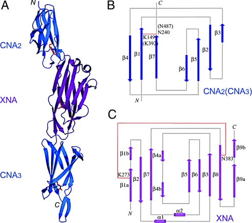
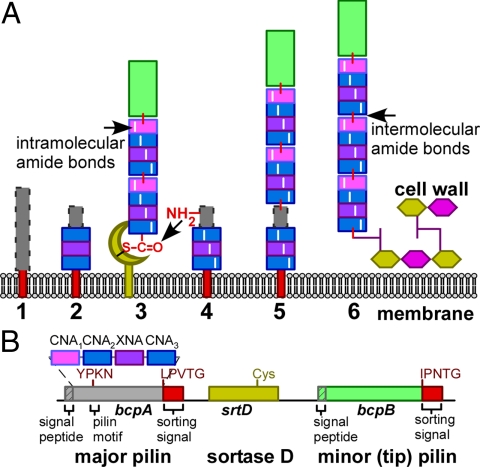
Gram-positive bacteria elaborate pili and do so without the participation of folding chaperones or disulfide bond catalysts. Sortases, enzymes that cut pilin precursors, form covalent bonds that link pilin subunits and assemble pili on the bacterial surface. We determined the x-ray structure of BcpA, the major pilin subunit of Bacillus cereus. The BcpA precursor encompasses 2 Ig folds (CNA(2) and CNA(3)) and one jelly-roll domain (XNA) each of which synthesizes a single intramolecular amide bond. A fourth amide bond, derived from the Ig fold of CNA(1), is formed only after pilin subunits have been incorporated into pili. We report that the domains of pilin precursors have evolved to synthesize a discrete sequence of intramolecular amide bonds, thereby conferring structural stability and protease resistance to pili.


Conflict of interest statement

The authors declare no conflict of interest.

Figures

Fig. 1.
Fig. 1.
Crystal structure of BcpA* and topology diagrams of the CNA and XNA domains. (A) The crystal structure of BcpA* was solved using MAD. The CNA2 and CNA3 domains are shown as blue ribbons and the XNA domain purple. The Lys and Asn residues forming the intramolecular amide bonds in each of the 3 crystallized domains are shown as red sticks. Topology diagrams of the CNA (B) and XNA (C) domains show the arrangement of β-strands in the reverse Ig-fold (CNA) and jelly-roll fold (XNA). Both CNA2 and CNA3 share the same overall topology. β-strands are shown as arrows and α-helices as cylinders. Intramolecular amide bonds are shown as red lines.
Fig. 2.
Fig. 2.
Four intramolecular and one intermolecular amide bond are formed in BcpA pili. (A) Schematic to illustrate the structure or BcpA pili assembled from the CNA1, CNA2, XNA, and CNA3 domains, the side chain residues (Lys and Asn) engaged in the formation of intramolecular amide bonds as well as the intermolecular amide bond between Lys162 (YPKN motif) and Thr522 (in the LPVT remnant of the cleaved sorting signal). BcpA pilin variants encoded by pJB232 harbor 6 internal methionine residues that can be cleaved with cyanogen bromide (CNBr) to generate homoserine lactone breaks. (B) B. anthracis srtA::ermC (pJB232) was stained with α-BcpA antibodies, 10 nm gold anti-rabbit conjugate, and uranyl acetate. Images were viewed at 8,260× and the bar represents a distance of 1 μm. (C) Pili were isolated from bacilli, cleaved with CNBr, purified by Ni-NTA chromatography and eluate analyzed by Coomassie stained SDS/PAGE. (D) The mass of compound B (arrow in C) was determined by LC-ESI-FTMS. (E) The structure of compound B as derived by Edman degradation and mass spectrometry. Diagram displays the peptide sequence of 6 branched peptides, generated by CNBr cleavage at 6 internal methionine (M) residues, and the intra- and intermolecular amide bonds that connect all 6 peptides.
Fig. 3.
Fig. 3.
Intramolecular amide bonds are required for the functional assembly of BcpA pili. (A) Diagram of polymerized BcpA pili with 4 intramolecular and one intermolecular amide bonds. (B–K) Wild-type BcpA pili and their variants were purified from B. anthracis srtA::ermC. Isolated pili were either mock treated or incubated with trypsin. The integrity of isolated pili was analyzed by immunogold labeling and transmission electron microscopy. See Table 1 for quantification of pilus length.
Fig. 4.
Fig. 4.
Model for pilus assembly in Bacillus cereus. (A) The unfolded BcpA precursor (1) is secreted across the plasma membrane and its N-terminal signal peptide removed. The CNA2, CNA3 and XNA domains assume reverse Ig or jelly-roll folds (2), form intramolecular amide bonds and enable presentation of the YPKN amino group (4) for nucleophilic attack at the sortase D-acyl intermediate with nascent pili, including the BcpB tip pilin (3). The product of this reaction initially links the incoming BcpA subunit to the LPXT motif of the growing pilus. At this stage, the CNA1 domain of the incoming BcpA subunit is still unfolded (gray box with dashed borders) (5). Following folding and intramolecular amide bond formation by the CNA1 domain, the mature, protease resistant pilus is immobilized in the cell wall envelope of bacilli (6). (B) Diagram displays the bcpA-srtD-bcpB gene cluster of B. cereus and the relevant features of the encoded products.

References

    1. Marraffini LA, DeDent AC, Schneewind O. Sortases and the art of anchoring proteins to the envelopes of gram-positive bacteria. Microbiol Mol Biol Rev. 2006;70:192–221. - PMC - PubMed
    1. Sarvas M, Harwood CR, Bron S, van Dijl JM. Post-translational folding of secretory proteins in Gram-positive bacteria. Biochim Biophys Acta. 2004;1694:311–327. - PubMed
    1. Ton-That H, Liu G, Mazmanian SK, Faull KF, Schneewind O. Purification and characterization of sortase, the transpeptidase that cleaves surface proteins of Staphylococcus aureus at the LPXTG motif. Proc Natl Acad Sci USA. 1999;96:12424–12429. - PMC - PubMed
    1. Budzik JM, et al. Amide bonds assemble pili on the surface of bacilli. Proc Natl Acad Sci USA. 2008;105:10215–10220. - PMC - PubMed
    1. Budzik JM, Oh SY, Schneewind O. Cell wall anchor structure of BcpA pili in Bacillus anthracis. J Biol Chem. 2008;283:36676–36686. - PMC - PubMed

Publication types

MeSH terms

LinkOut - more resources