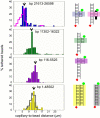
Single molecule detection of direct, homologous, DNA/DNA pairing
- PMID: 19903884
- PMCID: PMC2775704
- DOI: 10.1073/pnas.0911214106
Single molecule detection of direct, homologous, DNA/DNA pairing
Abstract
Using a parallel single molecule magnetic tweezers assay we demonstrate homologous pairing of two double-stranded (ds) DNA molecules in the absence of proteins, divalent metal ions, crowding agents, or free DNA ends. Pairing is accurate and rapid under physiological conditions of temperature and monovalent salt, even at DNA molecule concentrations orders of magnitude below those found in vivo, and in the presence of a large excess of nonspecific competitor DNA. Crowding agents further increase the reaction rate. Pairing is readily detected between regions of homology of 5 kb or more. Detected pairs are stable against thermal forces and shear forces up to 10 pN. These results strongly suggest that direct recognition of homology between chemically intact B-DNA molecules should be possible in vivo. The robustness of the observed signal raises the possibility that pairing might even be the "default" option, limited to desired situations by specific features. Protein-independent homologous pairing of intact dsDNA has been predicted theoretically, but further studies are needed to determine whether existing theories fit sequence length, temperature, and salt dependencies described here.
Conflict of interest statement
The authors declare no conflict of interest.
Figures


Similar articles
-
Early stages in RecA protein-catalyzed pairing. Analysis of coaggregate formation and non-homologous DNA contacts.J Mol Biol. 1992 Nov 20;228(2):409-20. doi: 10.1016/0022-2836(92)90830-d. J Mol Biol. 1992. PMID: 1453452
-
Single molecule identification of homology-dependent interactions between long ssRNA and dsDNA.Nucleic Acids Res. 2017 Jan 25;45(2):894-901. doi: 10.1093/nar/gkw758. Epub 2016 Aug 31. Nucleic Acids Res. 2017. PMID: 27580717 Free PMC article.
-
Statistical mechanics of homologous pairing of long double-stranded DNA.J Chem Phys. 2025 Jun 7;162(21):215102. doi: 10.1063/5.0265409. J Chem Phys. 2025. PMID: 40459361
-
Direct Homologous dsDNA-dsDNA Pairing: How, Where, and Why?J Mol Biol. 2020 Feb 7;432(3):737-744. doi: 10.1016/j.jmb.2019.11.005. Epub 2019 Nov 11. J Mol Biol. 2020. PMID: 31726060 Review.
-
Homologous genetic recombination as an intrinsic dynamic property of a DNA structure induced by RecA/Rad51-family proteins: a possible advantage of DNA over RNA as genomic material.Proc Natl Acad Sci U S A. 2001 Jul 17;98(15):8425-32. doi: 10.1073/pnas.111005198. Proc Natl Acad Sci U S A. 2001. PMID: 11459985 Free PMC article. Review.
Cited by
-
Homologous pairing in short double-stranded DNA-grafted colloidal microspheres.Biophys J. 2022 Dec 20;121(24):4819-4829. doi: 10.1016/j.bpj.2022.09.037. Epub 2022 Oct 4. Biophys J. 2022. PMID: 36196058 Free PMC article.
-
The 3D folding of metazoan genomes correlates with the association of similar repetitive elements.Nucleic Acids Res. 2016 Jan 8;44(1):245-55. doi: 10.1093/nar/gkv1292. Epub 2015 Nov 24. Nucleic Acids Res. 2016. PMID: 26609133 Free PMC article.
-
Recombination-independent recognition of DNA homology for repeat-induced point mutation.Curr Genet. 2017 Jun;63(3):389-400. doi: 10.1007/s00294-016-0649-4. Epub 2016 Sep 14. Curr Genet. 2017. PMID: 27628707 Free PMC article. Review.
-
Quantitative modeling of forces in electromagnetic tweezers.J Appl Phys. 2010 Nov 15;108(10):104701. doi: 10.1063/1.3510481. Epub 2010 Nov 18. J Appl Phys. 2010. PMID: 21258580 Free PMC article.
-
The emerging sequence grammar of 3D genome organisation.Hum Genet. 2025 Aug 25. doi: 10.1007/s00439-025-02772-8. Online ahead of print. Hum Genet. 2025. PMID: 40853480 Review.
References
-
- McKee BD. In: Meiosis. Benavente R, Volff J-N, editors. Basel: Karger; 2009. pp. 56–68.
-
- Weiner BM, Kleckner N. Chromosome pairing via multiple interstitial interactions before and during meiosis in yeast. Cell. 1994;77:977–991. - PubMed
-
- Keeney S, Kleckner N. Communication between homologous chromosomes: Genetic alterations at a nuclease-hypersensitive site can alter mitotic chromatin structure at that site both in cis and in trans. Genes Cells. 1996;1:475–489. - PubMed
Publication types
MeSH terms
Substances
Grants and funding
LinkOut - more resources
Full Text Sources
Other Literature Sources

