. 2009 Nov 24;106(47):20093-6.
doi: 10.1073/pnas.0904036106. Epub 2009 Nov 10.

Two size-selective mechanisms specifically trap bacteria-sized food particles in Caenorhabditis elegans

Affiliations

Two size-selective mechanisms specifically trap bacteria-sized food particles in Caenorhabditis elegans

Christopher Fang-Yen et al. Proc Natl Acad Sci U S A. .

Abstract

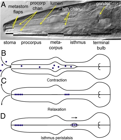
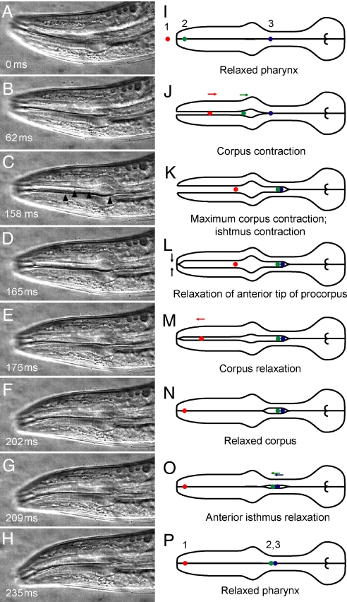
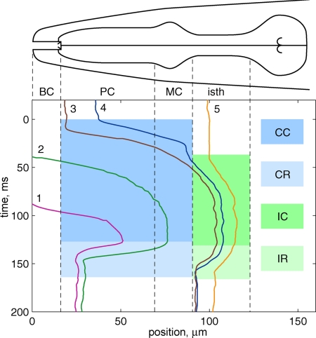
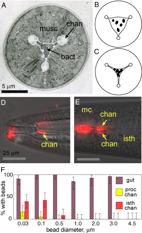
Caenorhabditis elegans is a filter feeder: it draws bacteria suspended in liquid into its pharynx, traps the bacteria, and ejects the liquid. How pharyngeal pumping simultaneously transports and filters food particles has been poorly understood. Here, we use high-speed video microscopy to define the detailed workings of pharyngeal mechanics. The buccal cavity and metastomal flaps regulate the flow of dense bacterial suspensions and exclude excessively large particles from entering the pharynx. A complex sequence of contractions and relaxations transports food particles in two successive trap stages before passage into the terminal bulb and intestine. Filtering occurs at each trap as bacteria are concentrated in the central lumen while fluids are expelled radially through three apical channels. Experiments with microspheres show that the C. elegans pharynx, in combination with the buccal cavity, is tuned to specifically catch and transport particles of a size range corresponding to most soil bacteria.


Conflict of interest statement

The authors declare no conflict of interest.

Figures

Fig. 1.
Fig. 1.
Pharyngeal anatomy and behavior. (A) Annotated DIC image of anterior of adult worm (field of view, 177 μm × 57 μm). The corpus includes the procorpus and metacorpus. Intestine is posterior to the terminal bulb. (B and C) Pharyngeal pumping. Contraction of pharyngeal muscle draws fluid containing suspended food particles (dots) into pharyngeal lumen. Relaxation ejects fluids while trapping particles. Curved arrows indicate flow of fluids. (D) Isthmus peristalsis carries food from anterior isthmus to terminal bulb and intestine.
Fig. 2.
Fig. 2.
High-speed video imaging of pharyngeal pumping. (A–H) Frames from high-speed video sequence showing movements of pharyngeal muscles and 0.75-μm polystyrene particles during pharyngeal pumping in adult worm (field of view, 136 μm × 65 μm). Black triangles in C indicate several particles. (I–P) Corresponding illustrations showing initial, intermediate, and final positions of three representative particles labeled 1 (red), 2 (green), and 3 (blue). During one cycle of contraction and relaxation, particle 1 has been transported from outside the pharynx to the anterior procorpus trap. Particle 2 has been transported from the anterior procorpus trap to the anterior isthmus trap. Particle 3 remains in the anterior isthmus trap. Full video sequence is available in Movie S1.
Fig. 3.
Fig. 3.
Trajectories of five bacteria during one pharyngeal pump cycle in an adult worm. BC, buccal cavity; PC, procorpus; MC, metacorpus; isth, isthmus. Shaded regions indicate timing of different pumping phases. CC, corpus contraction (0–127 ms); CR, corpus relaxation (127–164 ms); IC, isthmus contraction (36–132 ms); IR, isthmus relaxation (132–166 ms). Positions of bacteria are measured along approximate centerline of pharynx with zero at tip of nose (see Materials and Methods). Time denoted relative to onset of corpus contraction. Pumping period for this video sequence was t = 200 ms. Corresponding video can be found in Movie S4.
Fig. 4.
Fig. 4.
Particle filtering. (A) Electron micrograph cross-section of pharynx anterior procorpus containing bacteria. musc, muscles; chan, channels; bact, trapped bacteria. (B) Illustration of cross-section of contracted pharynx containing bacterial suspension. (C) Illustration of cross-section after relaxation. Bacteria are trapped; fluid has flowed radially into channels. (D) Overlay of DIC image and fluorescence image from worm incubated with 0.03-μm-diameter red fluorescent beads. Beads aggregated in procorpus channels. Ventral channel (arrow) shown; subdorsal channels are out of focus in this image. Beads also present at anterior tip of worm. (E) Overlay of DIC image and fluorescence image from worm incubated with 0.1-μm-diameter red fluorescent beads. mc, metacorpus; isth, isthmus. Beads have aggregated in the anterior isthmus channels (arrows) and in the pharyngeal lumen. (F) Size-dependent accumulation of fluorescent beads in pharyngeal channels and gut. Fraction of worms positive for beads in anterior procorpus channels, anterior isthmus channels, or intestinal lumen for various bead diameters. Number of worms n ≥ 33 for 0.03- to 1-μm beads, n ≥ 25 for 2- to 4.5-μm beads. Error bars indicate 95% confidence intervals for true mean.

Similar articles

Cited by

References

    1. Albertson DG, Thomson JN. The pharynx of Caenorhabditis elegans. Philos Trans R Soc Lond B Biol Sci. 1976;275:299–325. - PubMed
    1. Avery L. The genetics of feeding in Caenorhabditis elegans. Genetics. 1993;133:897–917. - PMC - PubMed
    1. Avery L, Shtonda BB. Food transport in the C elegans pharynx. J Exp Biol. 2003;206:2441–2457. - PMC - PubMed
    1. Purcell EM. Life at low Reynolds number. Am J Phys. 1977;45:3–11.
    1. Seymour MK, Wright KA, Doncaster CC. The action of the anterior feeding apparatus of Caenorhabditis elegans (Nematoda, Rhabditida) J Zool. 1983;201:527–539.

Publication types

LinkOut - more resources