. 2010 Jan 21;115(3):541-8.
doi: 10.1182/blood-2009-03-211383. Epub 2009 Nov 10.

The role of the Th1 transcription factor T-bet in a mouse model of immune-mediated bone-marrow failure

Affiliations

The role of the Th1 transcription factor T-bet in a mouse model of immune-mediated bone-marrow failure

Yong Tang et al. Blood. .

Abstract

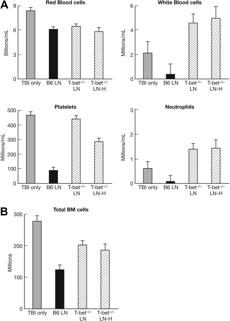
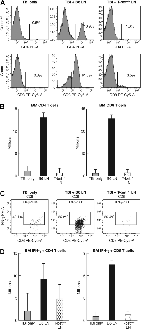
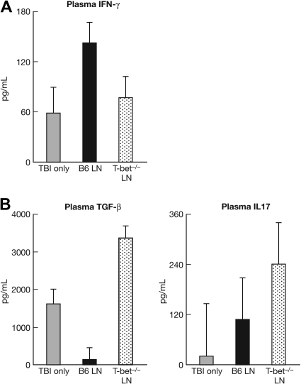
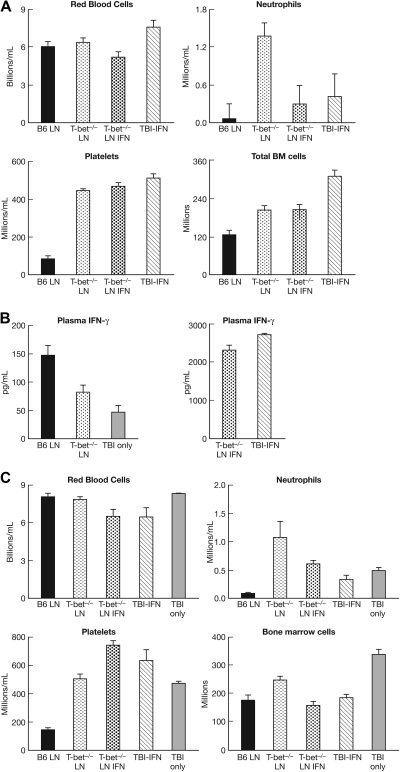
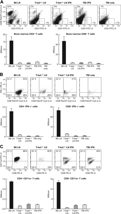
The transcription factor T-bet is a key regulator of type 1 immune responses. We examined the role of T-bet in an animal model of immune-mediated bone marrow (BM) failure using mice carrying a germline T-bet gene deletion (T-bet(-/-)). In comparison with normal C57BL6 (B6) control mice, T-bet(-/-) mice had normal cellular composition in lymphohematopoietic tissues, but T-bet(-/-) lymphocytes were functionally defective. Infusion of 5 x 10(6) T-bet(-/-) lymph node (LN) cells into sublethally irradiated, major histocompatibility complex-mismatched CByB6F1 (F1) recipients failed to induce the severe marrow hypoplasia and fatal pancytopenia that is produced by injection of similar numbers of B6 LN cells. Increasing T-bet(-/-) LN-cell dose to 10 to 23 x 10(6) per recipient led to only mild hematopoietic deficiency. Recipients of T-bet(-/-) LN cells had no expansion in T cells or interferon-gamma-producing T cells but showed a significant increase in Lin(-)Sca1(+)CD117(+)CD34(-) BM cells. Plasma transforming growth factor-beta and interleukin-17 concentrations were increased in T-bet(-/-) LN-cell recipients, possibly a compensatory up-regulation of the Th17 immune response. Continuous infusion of interferon-gamma resulted in hematopoietic suppression but did not cause T-bet(-/-) LN-cell expansion or BM destruction. Our data provided fresh evidence demonstrating a critical role of T-bet in immune-mediated BM failure.


Figures

Figure 1
Figure 1
T-bet−/− LN-cell malfunction in vivo in the induction of BM failure. Lymph node (LN) cells from normal B6 or T-bet−/− donors were used as effectors to induce bone marrow (BM) failure in sublethally irradiated (5 Gy) CByB6F1 recipient mice. Data were pooled from 4 different experiments showing as means with SEs for animals that received the following treatments: (1) TBI only (5 Gy TBI, n = 11), (2) B6 LN (5 Gy TBI + 5 × 10⁶ B6 LN, n = 13), (3) T-bet−/− LN (5 Gy TBI + 5 × 10⁶ T-bet−/− LN, n = 16), (4) T-bet−/− LN-H (5 Gy TBI + 10 × 106 T-bet−/− LN, n = 8). Total BM cells were calculated assuming that bilateral tibia and femurs contain 25% of total marrow cells. Infusion of B6 LN cells, but not T-bet−/− LN cells, caused significant decreases in WBCs (P < .01, panel A), neutrophils (P < .01, panel A), platelets (P < .01, panel A), and total BM cells (P < .01, panel B).
Figure 2
Figure 2
Impaired ability of T-bet−/− LN cells in the destruction of host hematopoietic stem and progenitor cells. BM cells from TBI-only (n = 11), B6 LN (n = 13), and T-bet−/− LN (n = 16) mice were stained and analyzed for the presence of hematopoietic stem and progenitor cells by use of the LinKit+Sca1+CD34 (KSLCD34) marker combination shown as representative dot plots (A) as well as means with SEs (B). Infusion of B6 LN cells reduced (P < .05) BM hematopoietic cells. As a surprise but consistent observation, infusion of T-bet−/− LN cells markedly increased (P < .01) the hematopoietic stem and progenitor cells in recipient BM.
Figure 3
Figure 3
T-cell expansion after the infusion of B6 and T-bet−/− LN cells. CByB6F1 mice that received TBI only (n = 11), B6 LN (n = 13), or T-bet−/− LN (n = 16) treatment were analyzed for CD4 and CD8 T-cell expansion in the BM shown as representative histograms (A) and means with SEs (B). There was drastic expansion (P < .01) of infused B6 T cells but not infused T-bet−/− T cells. We also analyzed the proportion of BM CD4 and CD8 T cells that express IFN-γ, shown as representatives (C) and means with SEs (D), because there was also significant expansion of IFN-γ–expressing CD4 (P < .05) and IFN-γ–expressing CD8 (P < .01) T cells in mice that received infusion of B6 LN cells but not T-bet−/− LN cells.
Figure 4
Figure 4
Up-regulation in Th17 immune response by T-bet−/− LN cell infusion. In 2 experiments, we measured plasma cytokine concentrations by enzyme-linked immunoassay as detailed in methods. Infusion of B6 LN cells (n = 6) resulted in greater (P < .05) plasma IFN-γ (A) concentration in comparison with TBI-only control mice. In contrast, infusion of T-bet−/− LN cells (n = 6) resulted in greater plasma TGF-β (P < .01, B) and IL17A (P > .05, B) concentrations relative to TBI-only control mice (n = 3).
Figure 5
Figure 5
Exogenous IFN-γ failure to rescue T-bet−/− LN-cell functional deficiency. In 2 experiments, we attempted to use exogenous IFN-γ to restore the ability for T-bet−/− LN cells to induce BM failure through intravenous and intraperitoneal injection. Injection of IFN-γ intraperitoneally once per day at 0.2 μg/mouse (6666 pg/g of body weight) from day 1 to day 11 to TBI-treated F1 recipients, with or without the infusion of 5 × 10⁶ T-bet−/− LN cells, reduced recipient neutrophils without showing any effect on red blood cells, platelets, or total BM cells (A). In 2 separate experiments, we infused T-bet−/− LN cells into sublethally irradiated CByB6F1 mice that were each installed with an osmotic pump to provide continuous infusion of IFN-γ as detailed in “Induction of BM failure.” After 12 days, plasma IFN-γ concentrations were drastically greater in mice that received IFN-γ infusion (B). Mice that received T-bet−/− LN cells and continuous IFN-γ infusion had lower levels of WBCs, neutrophils, red blood cells, and BM cells than did mice that had received T-bet−/− LN cells without IFN-γ (C). Data shown are means with SEs for each group: B6 LN (n = 7), T-bet−/− LN (n = 9), T-bet−/− LN IFN-γ (n = 9), TBI-IFN-γ (n = 4), and TBI only (n = 4).
Figure 6
Figure 6
Continuous IFN-γ infusion effects on T-cell expansion and activation. CD4 and CD8 T cells in the BM of recipient mice, as described in Figure 5, were measured by flow cytometry, shown as percentages in representative dotplots, and was calculated as total cells per animal, shown as means with SEs (A). Proportion and total number of CD8 T cells that expressed the inflammatory cytokine IFN-γ are also shown as representatives and means with SEs (B). T-cell activation was assessed by CD11a expression, shown in the same fashion (C).

Similar articles

Cited by

References

    1. Young NS, Scheinberg P, Calado RT. Aplastic anemia. Curr Opin Hematol. 2008;15(3):162–168. - PMC - PubMed
    1. Kojima S, Frickhofen N, Deeg HJ, et al. Aplastic anemia. Int J Hematol. 2005;82(5):408–411. - PubMed
    1. Nagy SM, Jr, Fisher JJ. Aplastic anemia and immunosuppression [abstract]. JAMA. 2003;290(2):193. - PubMed
    1. Abdelkefi A, Ben OT, Ladeb S, et al. Bone marrow transplantation for patients with acquired severe aplastic anemia using cyclophosphamide and antithymocyte globulin: the experience from a single center. Hematol J. 2003;4(3):208–213. - PubMed
    1. Maciejewski JP, Hibbs JR, Anderson S, Katevas P, Young NS. Bone marrow and peripheral blood lymphocyte phenotype in patients with bone marrow failure. Exp Hematol. 1994;22(11):1102–1110. - PubMed

MeSH terms