

Lipomatous pseudohypertrophy of the pancreas: a clinicopathologically distinct entity
- PMID: 19904221
- PMCID: PMC3164317
- DOI: 10.1097/MPA.0b013e3181bd2923
Lipomatous pseudohypertrophy of the pancreas: a clinicopathologically distinct entity
Abstract
Objectives: Owing to the challenges in obtaining pancreatic biopsies, pancreatic resection for presumed malignancy is often performed without histological confirmation. As a result, benign lesions are sometimes surgically removed. One such condition, which is poorly defined in the literature, is referred to as lipomatous pseudohypertrophy (LPH) of the pancreas.
Methods: Five cases of LPH were analyzed.
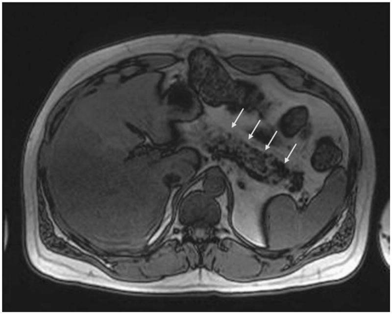
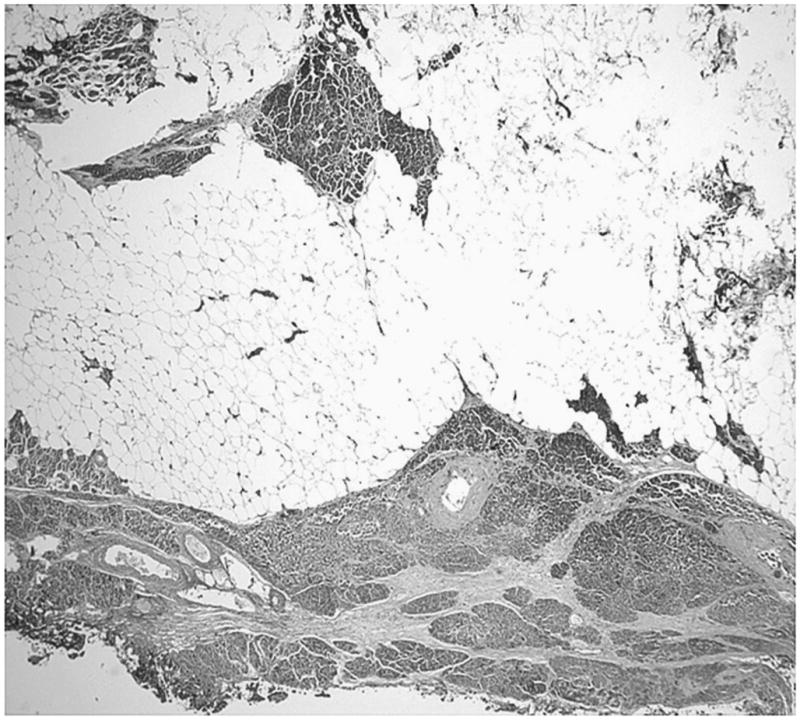
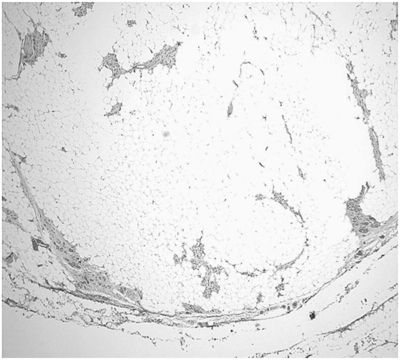
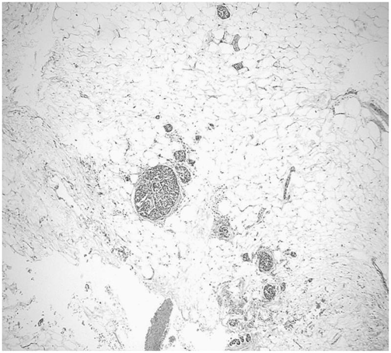
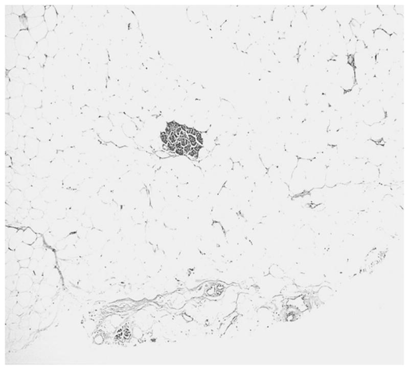
Results: Four patients underwent surgical resection, 3 of which were diagnosed preoperatively by radiology as having ductal adenocarcinoma. The fourth case was correctly interpreted by magnetic resonance imaging as LPH, but the patient underwent resection because of the intractable pain due to pancreatitis. The fifth patient has been placed on watchful waiting.Two tumors were in the pancreatic head, one in the tail, one in the uncinate process, and one demonstrated diffuse involvement. Microscopically, they were characterized as having normal lipocytes without lipoblasts or inflammation. Within the adipose tissue, scattered microscopic foci of pancreatic parenchyma could be seen.
Conclusion: Lipomatous pseudohypertrophy of the pancreas is a distinct entity characterized by localized/diffuse replacement of pancreatic parenchyma with mature adipose tissue. It forms a pseudotumor that may be difficult to distinguish clinically from pancreatic adenocarcinoma. This entity should be considered when evaluating patients with a new diagnosis of a hypodense pancreatic neoplasm on imaging.
Figures
References
-
- Kuroda N, Okada M, Toi M, et al. Lipomatous pseudohypertrophy of the pancreas: further evidence of advanced hepatic lesion as the pathogenesis. Pathol Int. 2003;53(2):98–101. - PubMed
-
- Sasaki M, Nakanuma Y, Ando H. Lipomatous pseudohypertrophy of the pancreas in a patient with cirrhosis due to chronic hepatitis B. Pathol Int. 1998;48(7):566–568. - PubMed
-
- Henkinbrant A, Khalek W, Farchakh E, et al. Cholestatic jaundice complicating lipomatous pseudo-hypertrophy of the pancreas [in French] Acta Gastroenterol Belg. 1990;53(3):315–322. - PubMed
-
- Flohr T, Bonatti H, Shumaker N, et al. Liver transplantation in a patient with primary sclerosing cholangitis suffering from lipomatous pseudohypertrophy of the pancreas. Transpl Int. 2008;21(1):89–91. - PubMed
Publication types
MeSH terms
Grants and funding
LinkOut - more resources
Full Text Sources
Medical
Research Materials
