. 2009 Dec 15;101(12):2023-9.
doi: 10.1038/sj.bjc.6605406. Epub 2009 Nov 10.

Self-production of tissue factor-coagulation factor VII complex by ovarian cancer cells

Affiliations

Self-production of tissue factor-coagulation factor VII complex by ovarian cancer cells

N Yokota et al. Br J Cancer. .

Abstract

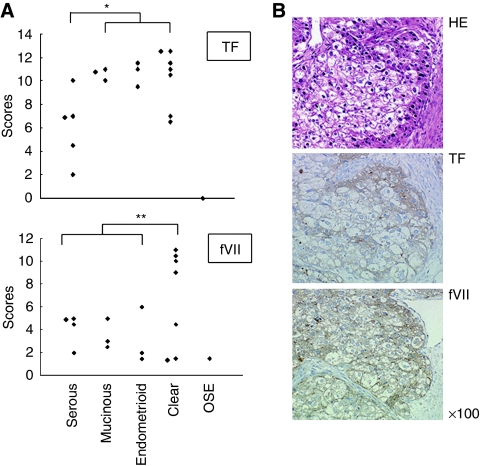
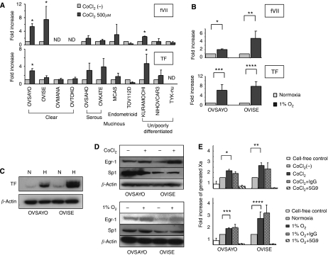
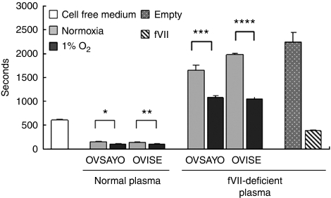
Background: Thromboembolic events are a major complication in ovarian cancer patients. Tissue factor (TF) is frequently overexpressed in ovarian cancer tissue and correlates with intravascular thrombosis. TF binds to coagulation factor VII (fVII), changing it to its active form, fVIIa. This leads to activation of the extrinsic coagulation cascade. fVII is produced by the liver and believed to be supplied from blood plasma at the site of coagulation. However, we recently showed that ovarian cancer cells express fVII transcripts under normoxia and that this transcription is inducible under hypoxia. These findings led us to hypothesise that ovarian cancer cells are intrinsically associated with TF-fVIIa coagulation activity, which could result in thrombosis.

Methods: In this study, we examined whether ectopically expressed fVII could cause thrombosis by means of immunohistochemistry, RT-PCR, western blotting and flow cytometry.

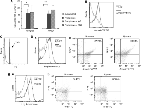
Results: Ectopic fVII expression occurs frequently in ovarian cancers, particularly in clear cell carcinoma. We further showed that ovarian cancer cells express TF-fVIIa on the cell surface under normoxia and that this procoagulant activity is enhanced by hypoxic stimuli. Moreover, we showed that ovarian cancer cells secrete microparticles (MPs) with TF-fVIIa activity. Production of this procoagulant secretion is enhanced under hypoxia.

Conclusion: These results raise the possibility that cancer cell-derived TF-fVIIa could cause thrombotic events in ovarian cancer patients.


Figures

Figure 1
Figure 1
Immunohistochemical analysis of TF and fVII expressions in ovarian cancer tissues. (A) Expression level of TF and fVII in ovarian cancer and normal ovarian surface epithelium (OSE) of a pre-menopausal ovary. Ovarian cancer tissues from serous, mucinous, endometrioid adenocarcinomas, clear cell carcinoma and OSE were stained with TF or fVII antibody. Expression levels were rated using the modified German immunoreactive score. *P=0.002, **P=0.039 (Student's t-test). (B) A representative example of immunohistochemical detection of TF and fVII in an ovarian clear cell carcinoma tissue that was scored as 11.5 and 11.0 for TF and fVII, respectively.
Figure 2
Figure 2
Real-time RT–PCR analysis of TF and fVII expressions in ovarian cancer cells. (A) Relative fVII and TF mRNA levels in ovarian cancer cells stimulated with 500 μM CoCl2 for 4 h. fVII transcription in OVSAYO and OVSAHO cells was reported earlier (Koizume et al, 2006) but was re-examined for comparison. *P<0.05, ND; not detected. (B) Relative increase of fVII and TF transcripts in OVSAYO and OVISE cells cultured under 1% O2 for 24 h compared with those cultured under normoxia (*P<0.001, **P=0.023, ***P=0.016, ****P=0.003). Columns for fVII and TF mRNA levels, mean (n=3); Bars, s.d. Data were statistically analysed using Student's t-test. (C) Western blotting analysis of TF levels in ovarian cancer cells. N and H are indicative of normoxia and hypoxia, respectively. (D) Western blotting analysis of Egr-1 and Sp1 expressions in ovarian cancer cells cultured under normoxia, hypoxia (1% O2 for 16 h) or hypoxia mimic (500 μM CoCl2 for 4 h) conditions. (E) Induction of procoagulant activity on the cell surface of ovarian cancer cells under CoCl2 or hypoxic conditions. Ovarian cancer cells were cultured under 500 μM CoCl2 for 11 h or under 1% O2 for 16 h, and then subjected to fXa generation assay. Columns for clotting time, mean (n=3); Bars, s.d. *P=0.001, **P=0.002, ***P<0.001, ****P=0.012.
Figure 3
Figure 3
Secretion of procoagulant activity by ovarian cancer cells. Ovarian cancer cells were cultured under normoxia or 1% O2 for 24 h, and then conditioned media were subjected to the plasma clotting assay using normal or fVII-deficient human plasma. FVII-containing cells and empty cells were indicative of stably fVII cDNA-transfected cells and stably transfected empty vector cells, respectively. Columns for clotting time, mean (n=3); Bars, s.d. *P=0.009, **P=0.003, ***P=0.002, ****P=0.0001.
Figure 4
Figure 4
Secretion of MPs with TF-fVIIa activity by ovarian cancer cells. (A) fXa generation activity of the supernatant and precipitate fraction of conditioned media prepared from OVSAYO and OVISE cells cultured under 1% O2. Procoagulant activity predominantly presented in the precipitate fraction and this activity was completely blocked by anti-TF (5G9) antibody treatment. *P<0.001, **P=0.001. (B) Flow cytometry analysis of precipitate fraction immunostained with PE-coupled anti-TF antibody and/or FITC-coupled annexin V showed that precipitate fraction prepared from conditioned media of OVSAYO cells was annexin-V positive. (C) The extracellular procoagulants analysed by flow cytometry. The grey peak corresponds to the 1-μm calibration microspheres. The white peak corresponds to the precipitate fraction prepared from conditioned media of OVSAYO cells. (D) Flow cytometry analysis of the precipitate fractions prepared from serum-free conditioned media of OVSAYO cells cultured under normoxia or 1% O2 for 24 h. (Da) The MP fraction prepared was TF positive. Background fluorescence was set with a fluorescent mouse IgG control. (Db) Dot plot of MP fraction immunostained with PE-coupled anti-TF antibody and FITC-coupled annexin V. Percentages shown in the upper right segment of the figure denote the relative amount of TF-positive MPs. (E) Flow cytometry analysis of the precipitate fractions prepared from serum-free conditioned media of OVSAYO cells cultured under normoxia or 1% O2 for 24 h. (Ea) The MP fraction prepared was fVII positive. Background fluorescence was set with a fluorescent mouse IgG control. (Eb) Dot plot of MP fraction immunostained with PE-coupled anti-TF antibody and FITC-coupled anti-fVII antibody. Percentages in the upper right segment of the figure denote amounts of fVII-positive fractions relative to total MPs.

References

    1. Amin C, Mackman N, Key NS (2008) Microparticles and cancer. Pathophysiol Haemost Thromb 36: 177–183 - PubMed
    1. Amirkhosravi A, Meyer T, Warnes G, Amaya M, Malik Z, Biggerstaff JP, Siddiqui FA, Sherman P, Francis JL (1998) Pentoxifylline inhibits hypoxia-induced upregulation of tumor cell tissue factor and vascular endothelial growth factor. Thromb Haemost 80: 598–602 - PubMed
    1. Chew HK, Wun T, Harvey DJ, Zhou H, White RH (2007) Incidence of venous thromboembolism and the impact on survival in breast cancer patients. J Clin Oncol 25: 70–76 - PubMed
    1. Davila M, Amirkhosravi A, Coll E, Desai H, Robles L, Colon J, Baker CH, Francis JL (2008) Tissue factor-bearing microparticles derived from tumor cells: impact on coagulation activation. J Thromb Haemost 6: 1517–1524 - PubMed
    1. Denko N, Schindler C, Koong A, Laderoute K, Green C, Giaccia A (2000) Epigenetic regulation of gene expression in cervical cancer cells by the tumor microenvironment. Clin Cancer Res 6: 480–487 - PubMed

Publication types