. 2010 Jul 1;127(1):32-42.
doi: 10.1002/ijc.25029.

Tumor inhibition by sodium selenite is associated with activation of c-Jun NH2-terminal kinase 1 and suppression of beta-catenin signaling

Affiliations

Tumor inhibition by sodium selenite is associated with activation of c-Jun NH2-terminal kinase 1 and suppression of beta-catenin signaling

Wenfeng Fang et al. Int J Cancer. .

Abstract

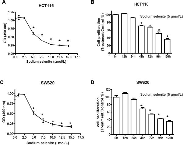
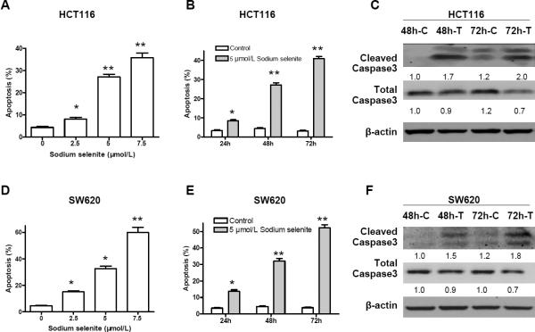
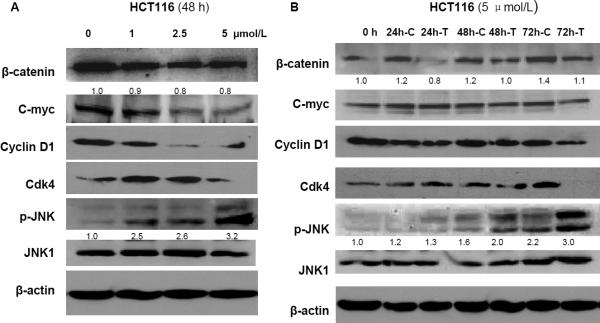
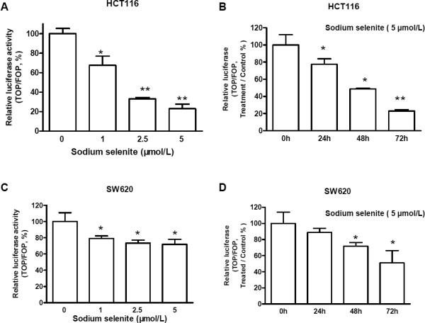
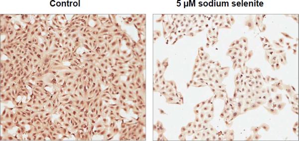
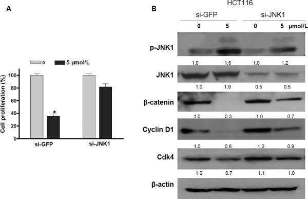
Epidemiological and clinical studies suggest that an increased intake of dietary selenium significantly reduces overall cancer risk, but the anticancer mechanism of selenium is not clear. In this study, we fed intestinal cancer mouse model. Muc2/p21 double mutant mice with a selenium-enriched (sodium selenite) diet for 12 or 24 weeks, and found that sodium selenite significantly inhibited intestinal tumor formation in these animals (p < 0.01), which was associated with phosphorylation of JNK1 and suppression of beta-catenin and COX2. In vitro studies showed that sodium selenite promoted cell apoptosis and inhibited cell proliferation in human colon cancer cell lines HCT116 and SW620. These effects were dose- and time course-dependent, and were also linked to an increase of JNK1 phosphorylation and suppression of beta-catenin signaling. Reduced JNK1 expression by small RNA interference abrogated sufficient activation of JNK1 by sodium selenite, leading to reduced inhibition of the beta-catenin signaling, resulting in reduced efficacy of inhibiting cell proliferation. Taken together, our data demonstrate that sodium selenite inhibits intestinal carcinogenesis in vivo and in vitro through activating JNK1 and suppressing beta-catenin signaling, a novel anticancer mechanism of selenium.


Figures

Figure 1
Figure 1
The Western-style diet (WD) accelerated mouse intestinal tumorigenesis, in comparison with the AIN-76A diet, but the Western-style diet supplemented with sodium selenite inhibited intestinal tumor formation in the Muc2−/−,p21+/− mice for 12 or 24 weeks (A), which occurred through activating JNK1 (JNK1 phosphorylation, p-JNK) and suppressing β-catenin and COX2 in intestinal epithelial cells from normal mucosa (B). β-actin was used as internal control. The image signal was quantified and normalized to β-actin. The ratio was presented. WD, western-style diet; WD+Sele, western-style diet supplemented with sodium selenite. N, number of animals studied in each group.
Figure 2
Figure 2
Sodium selenite inhibited cell proliferation in human colon cancer lines HCT116 and SW620. A and C, cell proliferation inhibition by sodium selenite occurred in a dosage dependent manner (48h); B and D, inhibition of cell proliferation by sodium selenite was time-dependent (5 μmol/L). * p<0.01, compared to the control group (0 μmol/L, or 0 h, respectively). These experiments were triplicated independently.
Figure 3
Figure 3
Sodium selenite promoted apoptosis in colon cancer lines HCT116 and SW620. A and D, apoptosis induction by sodium selenite was in a dosage dependent manner (48h); B and E, apoptosis induction by sodium selenite was time-dependent (5μmol/L); C and F, increase of cleaved Caspase 3, a biomarker of apoptosis, was observed in both colon cancer cell lines after 48 h and 72 h treatment of 5 μmol/L of sodium selenite, while the total caspases 3 was slightly reduced. “C” referred as “control”, and “T” referred as “sodium selenite treatment”. β-actin was used as loading control. The image signal was quantified and normalized to β- actin. The ratio was presented. These experiments were triplicated independently. (* p<0.05, ** p<0.01, compared to control, respectively).
Figure 4
Figure 4
Sodium selenite activated JNK1 (increase of phosphorylated JNK1, p-JNK1) and inhibited β-catenin signaling in dose- and time-dependent manner in colon cancer cells HCT116 (A and B) and SW620 (C and D), respectively. “C” referred as “control”, and “T” referred as “sodium selenite treatment”. β-actin was used as loading control. The image signal of p-JNK1 and β-catenin was quantified and normalized to β-actin. The ratio was presented. These experiments were triplicated independently.
Figure 4
Figure 4
Sodium selenite activated JNK1 (increase of phosphorylated JNK1, p-JNK1) and inhibited β-catenin signaling in dose- and time-dependent manner in colon cancer cells HCT116 (A and B) and SW620 (C and D), respectively. “C” referred as “control”, and “T” referred as “sodium selenite treatment”. β-actin was used as loading control. The image signal of p-JNK1 and β-catenin was quantified and normalized to β-actin. The ratio was presented. These experiments were triplicated independently.
Figure 5
Figure 5
Sodium selenite inhibited β-catenin/TCF4 transcriptional activities in dose- and time-dependent manner in colon cancer cells HCT116 (A and B) and SW620 (C and D). As described in “Materials and Methods”, colon cancer cells were co-transfected with TOPFlash (TOP) or FOPFlash (FOP) and with Renilla reporter plasmid using LipofectAMINE 2000. 6 h after transfection, the medium was removed and the cells were treated with fresh medium supplemented with 5 μmol/L of sodium selenite for 24 , 48 h and 72h, or treated with 0, 1, 2.5 ,5 μmol/L of sodium selenite for 48h. PBS was used as control. The presented data were the results from three independent luciferase activity assays (*p<0.05, **p<0.01, compared to control, respectively.)
Figure 6
Figure 6
Sodium selenite inhibited nuclear β-catenin expression as well as cytoplasmic expression in human osteosarcoma cells after 48 h treatment of 5 μmol/L of sodium selenite.
Figure 7
Figure 7
Cell growth inhibition by sodium selenite was JNK1 dependent in colon cancer cells HCT116. Knockdown of JNK1 by siRNA abrogated sodium selenite-mediated inhibition of cell growth (A) and suppression of the expression of β-catenin signaling pathway (B) (48 h). si-GFP was a small interfering RNA targeting human green fluorescence protein (GFP) and was used as mock control; si-JNK1 was a small interfering RNA targeting human JNK1. The knockdown efficiency was confirmed by western blotting. These experiments were triplicated independently. The image signal was quantified and normalized to β-actin. The ratio was presented right below the image. (* p<0.01, compared to the “0”)

Similar articles

Cited by

References

    1. Wang L, Bonorden MJ, Li GX, Lee HJ, Hu H, Zhang Y, Liao JD, Cleary MP, Lu J. Methyl-selenium compounds inhibit prostate carcinogenesis in the transgenic adenocarcinoma of mouse prostate model with survival benefit. Cancer Prev Res (Phila Pa) 2009;2:484–95. - PMC - PubMed
    1. Facompre N, El-Bayoumy K. Potential stages for prostate cancer prevention with selenium: implications for cancer survivors. Cancer Res. 2009;69:2699–703. - PMC - PubMed
    1. Clark LC, Combs GF, Jr., Turnbull BW, Slate EH, Chalker DK, Chow J, Davis LS, Glover RA, Graham GF, Gross EG, Krongrad A, Lesher JL, Jr., et al. Effects of selenium supplementation for cancer prevention in patients with carcinoma of the skin. A randomized controlled trial. Nutritional Prevention of Cancer Study Group. Jama. 1996;276:1957–63. - PubMed
    1. Greenwald P, Anderson D, Nelson SA, Taylor PR. Clinical trials of vitamin and mineral supplements for cancer prevention. Am J Clin Nutr. 2007;85:314S–7S. - PubMed
    1. Hawk ET, Levin B. Colorectal cancer prevention. J Clin Oncol. 2005;23:378–91. - PubMed

Publication types