. 2010 Jan;28(1):5-16.
doi: 10.1002/stem.254.

NOTCH pathway blockade depletes CD133-positive glioblastoma cells and inhibits growth of tumor neurospheres and xenografts

Affiliations

NOTCH pathway blockade depletes CD133-positive glioblastoma cells and inhibits growth of tumor neurospheres and xenografts

Xing Fan et al. Stem Cells. 2010 Jan.

Abstract

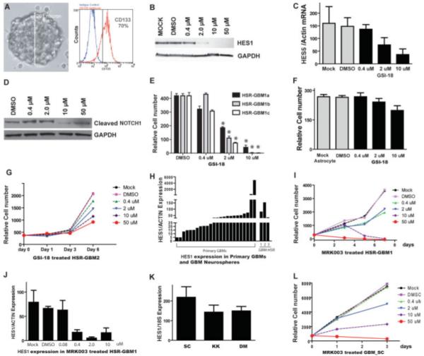
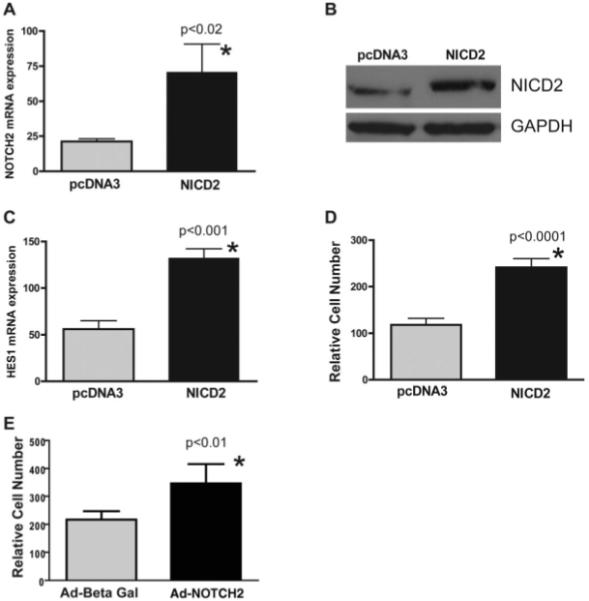
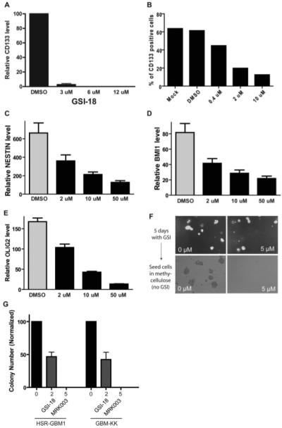
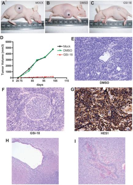
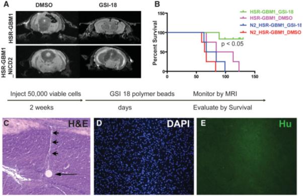
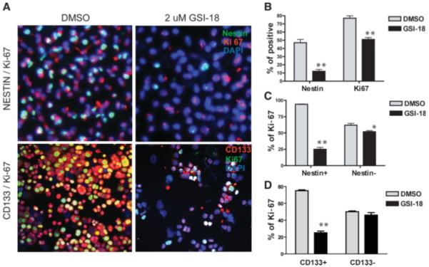
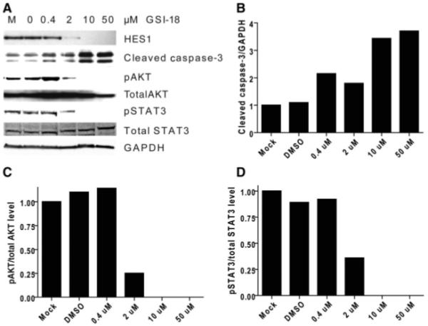
Cancer stem cells (CSCs) are thought to be critical for the engraftment and long-term growth of many tumors, including glioblastoma (GBM). The cells are at least partially spared by traditional chemotherapies and radiation therapies, and finding new treatments that can target CSCs may be critical for improving patient survival. It has been shown that the NOTCH signaling pathway regulates normal stem cells in the brain, and that GBMs contain stem-like cells with higher NOTCH activity. We therefore used low-passage and established GBM-derived neurosphere cultures to examine the overall requirement for NOTCH activity, and also examined the effects on tumor cells expressing stem cell markers. NOTCH blockade by gamma-secretase inhibitors (GSIs) reduced neurosphere growth and clonogenicity in vitro, whereas expression of an active form of NOTCH2 increased tumor growth. The putative CSC markers CD133, NESTIN, BMI1, and OLIG2 were reduced following NOTCH blockade. When equal numbers of viable cells pretreated with either vehicle (dimethyl sulfoxide) or GSI were injected subcutaneously into nude mice, the former always formed tumors, whereas the latter did not. In vivo delivery of GSI by implantation of drug-impregnated polymer beads also effectively blocked tumor growth, and significantly prolonged survival, albeit in a relatively small cohort of animals. We found that NOTCH pathway inhibition appears to deplete stem-like cancer cells through reduced proliferation and increased apoptosis associated with decreased AKT and STAT3 phosphorylation. In summary, we demonstrate that NOTCH pathway blockade depletes stem-like cells in GBMs, suggesting that GSIs may be useful as chemotherapeutic reagents to target CSCs in malignant gliomas.


Figures

Figure 1
Figure 1
GBM neurospheres are sensitive to NOTCH pathway blockade in vitro. (A): The GBM neurosphere line HSR-GBM1 grows as large, multicellular spheres. The majority (~70%) of neurosphere cells express CD133. (B): Upon GSI-18 treatment, protein levels of HES1, a NOTCH pathway target gene, were reduced after 48 hours of GSI-18 treatment of HSR-GBM1 neurospheres. (C, D): Upon GSI-18 treatment, mRNA expression of NOTCH target gene HES5 and protein levels of cleaved active NOTCH1 intracellular domain are also reduced. (E): GSI-18 reduced total cell mass in a dose-dependent fashion in all three HSR-GBM1 sublines (*, p < .05). (F): There is no significant change in normal human astrocyte growth when treated with GSI-18. (G): Growth of a second GBM neurosphere line, HSR-GBM2, was also inhibited by GSI-18. (H): HES1 mRNA levels fell into a similar range in primary GBM and tumor-derived neurospheres grown in culture. (I): A second γ-secretase inhibitor, MRK-003, could also suppress GBM neurosphere growth. (J): MRK003 showed potency similar to GSI-18 in terms of its ability to lower HES1 mRNA levels in tumor neurospheres. (K, L): Low-passage neurospheres GBM-SC, GBM-KK, and GBM-DM express the NOTCH target gene HEY1 and had their growth inhibited by NOTCH blockade. Abbreviations: DMSO, dimethyl sulfoxide; GAPDH, glyceraldehyde-3-phosphate dehydrogenase; GBM, glioblastoma; GSI-18, γ-secretase inhibitor 18.
Figure 2
Figure 2
Activation of NOTCH2 in glioblastoma (GBM) neurospheres increases tumor growth in vitro. (A): When the active form of NOTCH2 receptor (NICD2) was stably transfected into HSR-GBM1a cells, receptor mRNA levels were elevated. (B): NICD2 protein expression was also increased in NICD2 stably transfected HSR-GBM1 neurospheres detected by Western blot. GAPDH was used as protein loading control. (C): The NOTCH target gene HES1 was also induced in NICD2-transfected GBM neurospheres. (D): Cell growth was significantly increased in NICD2-transfected cells. (E): Transient expression of NICD2 in HSR-GBM1 neurospheres using adenovirus vectors also increased cell growth over 2 days. Abbreviations: Ad, Adenovirus; Beta Gal, β-galactosidase; GAPDH, glyceral-dehyde-3-phosphate dehydrogenase; NICD2, NOTCH2 intracellular domain.
Figure 3
Figure 3
NOTCH blockade depletes CD133-positive cells and inhibits neurosphere clonogenicity. (A): CD133 mRNA levels were severely decreased following GSI-18 treatment of HSR-GBM1 cells. (B): The percentage of CD133-positive cells in these cultures was also lowered by NOTCH blockade. (C–E): mRNA levels of the stem cell markers NESTIN, BMI1, and OLIG2 were also reduced in a dose-dependent fashion following GSI-18 treatment of HSR-GBM1 neurospheres. (F, G): Growing HSR-GBM1 cultures treated for 5 days with γ-secretase inhibitor can form some neurospheres (right top panel), but when equal numbers of DMSO- and MRK003-treated cells are seeded singly in methylcellulose, the latter cannot efficiently form colonies. Abbreviations: DMSO, dimethyl sulfoxide; GAPDH, glyceraldehyde-3-phosphate dehydrogenase; GBM, glioblastoma; GSI-18, γ-secretase inhibitor 18.
Figure 4
Figure 4
NOTCH signaling blockade depletes cells required for glioblastoma (GBM) propagation in vivo. (A–C): Large xenografts formed at all eight sites (asterisk) injected with cells in which NOTCH signaling was active, including nontreated (n = 4, A) and vehicle (DMSO)-treated (n = 4) cultures. Mice injected with equal number of viable cells previously treated with 2 μM GSI-18 (n = 6) were unable to efficiently form tumors, with small lesions developing at only three of six injection sites. (D): Tumor volume was dramatically reduced in mice engrafted with 2 μM GSI-18-pretreated cells compared with mice engrafted with mock or DMSO-pretreated cells. (E–G): The small subcutaneous xenografts that formed from cells pretreated with GSI-18 appeared similar to control tumors, and expressed abundant amounts of the NOTCH pathway target HES1 on immunohistochemical analysis. (H, I): Intracranial xenografts of HSR-GBM1 cells pretreated with DMSO or GSI-18 often invaded into the subventricular space (H), and occasionally formed necrotic foci with pseudopalisading (I). (Original magnifications both ×100.) Abbreviations: DMSO, dimethyl sulfoxide; GSI-18, γ-secretase inhibitor 18.
Figure 5
Figure 5
Local GSI treatment prolongs survival of mice bearing intracranial GBM xenografts. (A): Wild-type or NICD2 stably transfected GBM neurospheres were implanted into mouse brain, and GSI-18- or DMSO-soaked beads were injected into the tumor beds 2 weeks later. Tumor growth was monitored by MRI in selected animals, and large xenografts formed in DMSO- but not GSI-18-treated mice. As expected, constitutive NICD2 expression rendered xenografts insensitive to GSI-18. (B): GSI-18-treated mice have significantly prolonged survival compared with DMSO-treated mice, whereas expression of activated NOTCH2 promoted more rapid intracranial tumor growth. (C–E): Microscopic examination of a GSI-18-treated mouse brain stained with H&E reveals a round polymer bead (arrow) with a pale linear injection tract leading upward (arrowhead), but no tumor mass on this or additional step sections (original magnification ×100). Immunocytochemical stains using an antibody specific for human nuclei confirms the absence of viable tumor cells (D, E). (Original magnification ×100.) Abbreviations: DAPI, 4′,6-diamidino-2-phenylindole; DMSO, dimethyl sulfoxide; GAPDH, glyceraldehyde-3-phosphate dehydrogenase; GBM, glioblastoma; GSI-18, γ-secretase inhibitor 18; Hu, human nuclei; MRI, magnetic resonance imaging; NICD2, NOTCH2 intracellular domain.
Figure 6
Figure 6
NOTCH signaling regulates proliferation of poorly differentiated GBM cells. (A): NOTCH pathway blockade by GSI-18 reduces the NESTIN- or CD133-positive population in neurosphere lines, as assessed by double immunolabeling (upper panel: NESTIN in green; Ki-67, red; DAPI, blue; lower panel: CD133 in red; Ki-67, green; DAPI, blue). (B): Both NESTIN- and Ki-67-positive populations were significantly reduced. (C, D): Ki-67 proliferation indices were significantly lowered by GSI-18 in the NESTIN- or CD133-positive cell population, whereas NESTIN- or CD133-negative cells showed a lesser reduction (p value for t tests: *, p < .05, **, p < .01). Abbreviations: DAPI, 4′,6-diamidino-2-phenylindole; DMSO, dimethyl sulfoxide; GSI-18, γ-secretase inhibitor 18.
Figure 7
Figure 7
NOTCH signaling blockade promotes caspase-3 cleavage and reduces phosphorylation of AKT and STAT3. (A): Loss of AKT and STAT3 phosphorylation and activation of caspase-3 cleavage is induced by 2 μM or higher levels of GSI-18, whereas overall levels of AKT and STAT3 are unchanged in GBM neurosphere cells. GAPDH is used as a loading control. These changes were quantitated using densitometry in (B–D). Abbreviations: DMSO, dimethyl sulfoxide; GAPDH, glyceraldehyde-3-phosphate dehydrogenase; GBM, glioblastoma; GSI-18, γ-secretase inhibitor 18.

Similar articles

Cited by

References

    1. Reardon DA, Rich JN, Friedman HS, et al. Recent advances in the treatment of malignant astrocytoma. J Clin Oncol. 2006;24:1253–1265. - PubMed
    1. Louis DN, Ohgaki H, Wiestler OD, et al. WHO Classification of Tumours of the Central Nervous System. IARC Press; Lyon, France: 2007. - PMC - PubMed
    1. Stupp R, Mason WP, van den Bent MJ, et al. Radiotherapy plus concomitant and adjuvant temozolomide for glioblastoma. N Engl J Med. 2005;352:987–996. - PubMed
    1. Reya T, Morrison SJ, Clarke MF, et al. Stem cells, cancer, and cancer stem cells. Nature. 2001;414:105–111. - PubMed
    1. Hemmati HD, Nakano I, Lazareff JA, et al. Cancerous stem cells can arise from pediatric brain tumors. Proc Natl Acad Sci U S A. 2003;100:15178–15183. - PMC - PubMed

Publication types

MeSH terms