

Buckyballs meet viral nanoparticles: candidates for biomedicine
- PMID: 19904938
- PMCID: PMC2797550
- DOI: 10.1021/ja902293w
Buckyballs meet viral nanoparticles: candidates for biomedicine
Abstract
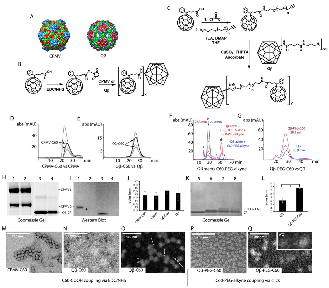
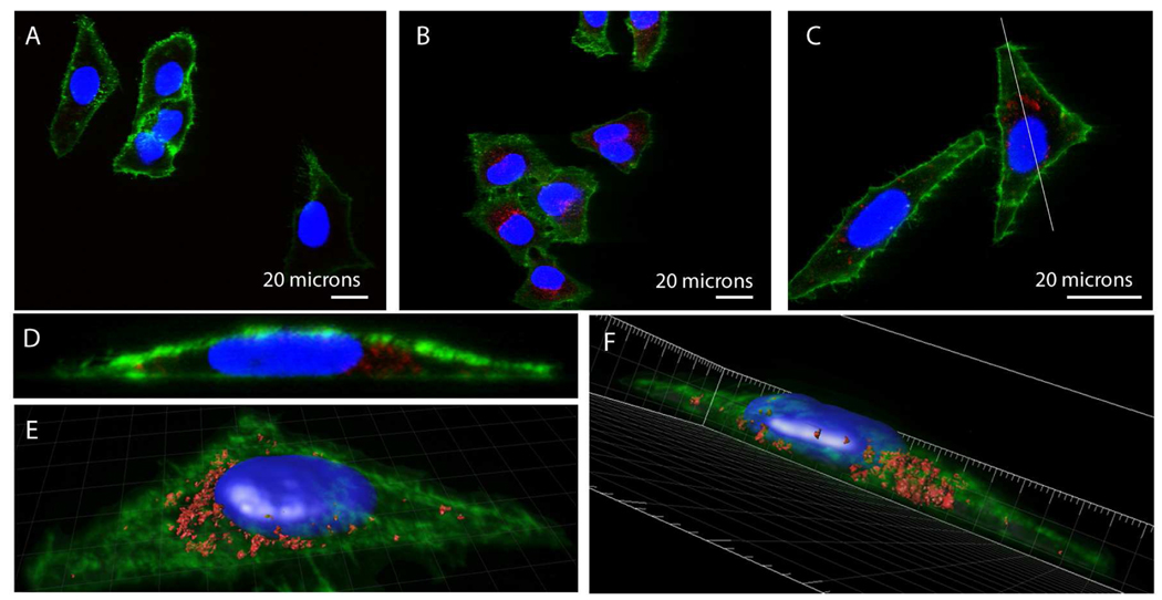
Fullerenes such as C(60) show promise as functional components in several emerging technologies. For biomedical applications, C(60) has been used in gene- and drug-delivery vectors, as imaging agents, and as photosensitizers in cancer therapy. A major drawback of C(60) for bioapplications is its insolubility in water. To overcome this limitation, we covalently attached C(60) derivatives to Cowpea mosaic virus and bacteriophage Qbeta virus-like particles, which are examples of naturally occurring viral nanoparticle (VNP) structures that have been shown to be promising candidates for biomedicine. Two different labeling strategies were employed, giving rise to water-soluble, stable VNP-C(60) and VNP-PEG-C(60) conjugates. Samples were characterized using a combination of transmission electron microscopy, scanning transmission electron microscopy (STEM), gel electrophoresis, size-exclusion chromatography, dynamic light scattering, and Western blotting. "Click" chemistry bioconjugation using a poly(ethylene glycol) (PEG)-modified propargyl-O-PEG-C(60) derivative gave rise to high loadings of fullerene on the VNP surface, as indicated by the imaging of individual C(60) units using STEM. The cellular uptake of dye-labeled VNP-PEG-C(60) complexes in a human cancer cell line was found by confocal microscopy to be robust, showing that cell internalization was not inhibited by the attached C(60) units. These results open the door for the development of novel therapeutic devices with potential applications in photoactivated tumor therapy.
Figures
References
-
- Thompson BC, Frechet JM. Angew Chem Int Ed Engl. 2008;47:58–77. - PubMed
- Taylor R, Walton D. Nature. 1993;363:685–693.
-
- Mroz P, Pawlak A, Satti M, Lee H, Wharton T, Gali H, Sarna T, Hamblin MR. Free Radic Biol Med. 2007;43:711–719. - PMC - PubMed
- Qu X, Komatsu T, Sato T, Glatter O, Horinouchi H, Kobayashi K, Tsuchida E. Bioconjugate Chem. 2008;19:1556–1560. - PubMed
- Zhao B, He YY, Bilski PJ, Chignell CF. Chem Res Toxicol. 2008;21:1056–1063. - PMC - PubMed
-
- Andersson T, Westman G, Wennerstroem O, Sundhahl M. J. Chem. Soc. Perkin Trans. 1994;2:1097–1101.
- Hummelen J, Knight B, LePeq F, Wudl F. J. Org. Chem. 1995;60:532–538.
- Sijbesma R, Srdanov G, Wudl F, Castoro J, Wilkins C, Friedman S, DeCamp D, Kenyon G. J.Am Chem. Soc. 1993;115:6510–6512.
- Sitharaman B, Zakharian T, Saraf A, Misra P, Ashcroft J, Pan S, Pham Q, Mikos A, Wilson L, Engler D. Mol. Pharm. 2008;5:567–578. - PMC - PubMed
- Vasella A, Uhlmann P, Waldruff C, Diederich F, Thilgen C. Angew. Chem, Int, Ed. 1992;31:1388–1390.
-
- Manchester M, Steinmetz NF. Viruses and Nanotechnology. Vol. 327. Berlin Heidelberg: Springer Verlag; 2008.
- Young M, Willits D, Uchida M, Douglas T. Annu Rev Phytopathol. 2008;46:361–384. - PubMed
Publication types
MeSH terms
Grants and funding
LinkOut - more resources
Full Text Sources
Other Literature Sources
