. 2009 Dec 8;48(48):11370-80.
doi: 10.1021/bi901368e.

The 8 and 5 kDa fragments of plasma gelsolin form amyloid fibrils by a nucleated polymerization mechanism, while the 68 kDa fragment is not amyloidogenic

Affiliations

The 8 and 5 kDa fragments of plasma gelsolin form amyloid fibrils by a nucleated polymerization mechanism, while the 68 kDa fragment is not amyloidogenic

James P Solomon et al. Biochemistry. .

Abstract

Familial amyloidosis of Finnish type (FAF), or gelsolin amyloidosis, is a systemic amyloid disease caused by a mutation (D187N/Y) in domain 2 of human plasma gelsolin, resulting in domain 2 misfolding within the secretory pathway. When D187N/Y gelsolin passes through the Golgi, furin endoproteolysis within domain 2 occurs as a consequence of the abnormal conformations that enable furin to bind and cleave, resulting in the secretion of a 68 kDa C-terminal fragment (amino acids 173-755, C68). The C68 fragment is cleaved upon secretion from the cell by membrane type 1 matrix metalloprotease (MT1-MMP), affording the 8 and 5 kDa fragments (amino acids 173-242 and 173-225, respectively) comprising the amyloid fibrils in FAF patients. Herein, we show that the 8 and 5 kDa gelsolin fragments form amyloid fibrils by a nucleated polymerization mechanism. In addition to demonstrating the expected concentration dependence of a nucleated polymerization reaction, the addition of preformed amyloid fibrils, or "seeds", was shown to bypass the requirement for the formation of a high-energy nucleus, accelerating 8 and 5 kDa D187N gelsolin amyloidogenesis. The C68 fragment can form small oligomers, but not amyloid fibrils, even when seeded with preformed 8 kDa fragment plasma gelsolin fibrils. Because the 68 kDa fragment of gelsolin does not form amyloid fibrils in vitro or in a recently published transgenic mouse model of FAF, we propose that administration of an MT1-MMP inhibitor could be an effective strategy for the treatment of FAF.


Figures
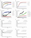
Figure 1
Figure 1
Relative amyloidogenicity of the 68, 8, and 5 kDa plasma gelsolin fragments (corresponding to amino acids 173–755, 173–242, and 173–225, respectively). Aggregation was monitored by the increase in TfT fluorescence in the plate reader TfT aggregation assay (see experimental procedures for details). The t50 associated with each curve is defined as the time required to reach half the maximum fluorescence and is shown in the legends of A and D. A. Freshly monomerized 8 kDa fragment (8 µM) was assayed at pHs varying from 6.0 to 7.5. B–C. Plasma gelsolin and fragments thereof (4 µM) were examined by the TfT fluorescence plate reader assay at pH 6.5 (B) and pH 6.8 (C). D. Comparison of the amyloidogenicity of the 8 and 5 kDa fragments at higher concentrations (pH 7.4). E–H. Various conditions known to promote amyloid fibril formation were applied to C68 (1.5 µM ) at pH 6.5. E. Buffer alone, control series as a reference for F–H. F. Heparin (10 µg/mL) was added to the C68 solution. G. Preformed 8 kDa fibrils were sonicated to generate fibrils of uniform length which were then added to the C68 solution. H. Guanidine HCl (0.5 M) was added to the C68 solution.
Figure 2
Figure 2
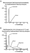
C68 was examined by analytical ultracentrifugation (AUC) to quantify the presence of monomer, dimer, trimer, or higher order oligomeric species. Values for integration of the peaks are given in the text. A. Freshly monomerized C68 (3 µM) exhibits one major peak corresponding to a monomer. B. Monomerized C68 (3 µM) was subjected to overhead rotation (24 rpm) for 16 h before analysis by AUC velocity experiments to demonstrate the presence of higher order species in addition to the monomer peak.
Figure 3
Figure 3
Concentration dependence of 8 kDa plasma gelsolin fragment amyloidogenesis analyzed at pH 7.4 using the TfT plate reader assay. Concentrations of the 8 kDa fragment ranged from 2 to 48 µM, as indicated. The t50 for each curve is defined as the time required to reach half the fluorescence at which the fibril extension reaction is completed and is shown in the legend.
Figure 4
Figure 4
The addition of preformed amyloid fibrils or “seeds” accelerates 8 and 5 kDa gelsolin fragment amyloidogenesis. In each experiment, preformed fibrils were sonicated for 20 min to generate fibrils of uniform length, a known quantity of which were added to the solution at the start of the plate reader TfT fluorescence aggregation assay. The t50 for each curve is defined as the time required to reach half the fluorescence of the fibril extension reaction. The t50 values are shown in the legends. A. Amyloidogenesis of the monomerized 8 kDa gelsolin fragment in the presence of increasing concentrations of preformed 8 kDa fibrils at pH 7.4. The final concentration of 8 kDa gelsolin fragment was 16 µM, including the added seed. B. The addition of 10% 8 kDa seed was compared to the addition of 10% 5 kDa seed to monomerized 8 kDa gelsolin fragment (14.4 µM, to give a final concentration of gelsolin fragment of 16 µM). The pH for this experiment was 6.5. C and D. Amyloidogenesis of the monomerized 5 kDa gelsolin fragment in the presence of various concentrations of preformed 5 kDa fibril seeds at pH 6.5 (C) and preformed 8 kDa fibril seeds at pH 7.0 (D). The final concentration of gelsolin fragment(s) were 16 µM, including the seed.
Figure 5
Figure 5
A freshly monomerized 8 kDa plasma gelsolin fragment solution (16 µM) was mixed at pH 7.0 by overhead rotation (24 rpm) at 37 °C. A. At various times indicated on the time course, an aliquot was removed and the TfT fluorescence was measured. At the three time points indicated by filled red squares, (4h, 12h, and 64h), the morphology of the fibrils were examined by atomic force microscopy (AFM). Each AFM image is a scan of a 5µm × 5µm area of the mica. B. AFM after the solution was mixed for 4 h (first red square), showing the presence of small fibrils and oligomers. C. AFM after the solution was mixed for 12 h (second red square) showing fully formed fibrils. D. After continued mixing for 64 h (third red square), the AFM shows laterally associated fibrils.

Similar articles

Cited by

References

    1. Kelly JW. Alternative conformations of amyloidogenic proteins govern their behavior. Curr. Opin. Struct. Biol. 1996;6:11–17. - PubMed
    1. Kelly JW. The alternative conformations of amyloidogenic proteins and their multi-step assembly pathways. Curr. Opin. Struct. Biol. 1998;8:101–106. - PubMed
    1. Selkoe DJ. Folding proteins in fatal ways. Nature. 2003;426:900–904. - PubMed
    1. Dobson CM. Protein folding and misfolding. Nature. 2003;426:884–890. - PubMed
    1. Cohen FE, Kelly JW. Therapeutic approaches to protein-misfolding diseases. Nature. 2003;426:905–909. - PubMed

Publication types