Comparative Study
. 2009 Dec 15;48(49):11786-95.
doi: 10.1021/bi9012947.

Phosphorylation of serine 205 by the protein kinase CK2 persists on Pax3-FOXO1, but not Pax3, throughout early myogenic differentiation

Affiliations
Comparative Study

Phosphorylation of serine 205 by the protein kinase CK2 persists on Pax3-FOXO1, but not Pax3, throughout early myogenic differentiation

Kevin N Dietz et al. Biochemistry. .

Abstract

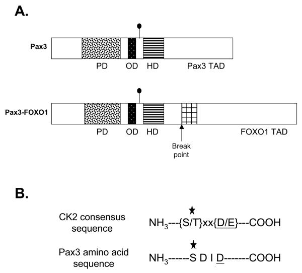
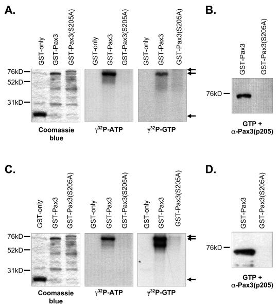
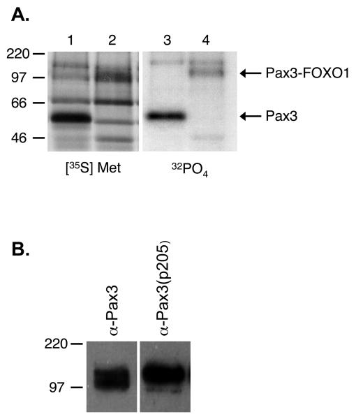
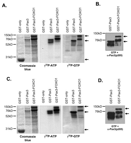
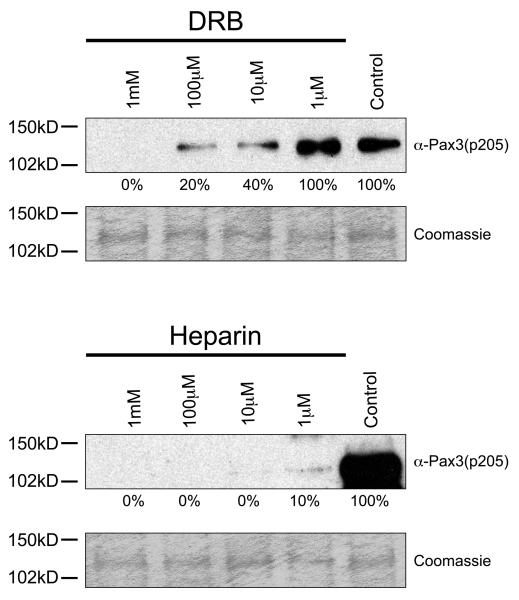
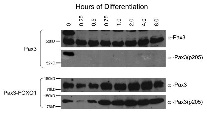
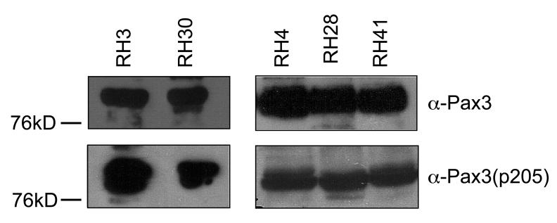
The myogenic transcription factor Pax3 plays an essential role in early skeletal muscle development and is a key component in alveolar rhabdomyosarcoma (ARMS), a childhood solid muscle tumor. ARMS is characterized by a t(2;13) chromosomal translocation resulting in the fusion of the 5' Pax3 sequences to the 3' FOXO1 sequences to encode the oncogenic fusion protein, Pax3-FOXO1. Posttranslational modifications such as phosphorylation are common mechanisms by which transcription factors are regulated. Consistent with this fact, we demonstrated in a previous report that Pax3 is phosphorylated on Ser205 in proliferating, but not differentiated, primary myoblasts. However, the kinase that mediates this phosphorylation event has yet to be identified. In addition, it is not known whether Pax3-FOXO1 is phosphorylated at this site or how the phosphorylation of the fusion protein changes during early myogenic differentiation. In this report we identify CK2 (formerly termed "casein kinase II") as the kinase responsible for phosphorylating Pax3 and Pax3-FOXO1 at Ser205 in proliferating mouse primary myoblasts. Furthermore, we demonstrate that, in contrast to wild-type Pax3, phosphorylation at Ser205 persists on Pax3-FOXO1 throughout early myogenic differentiation. Finally, we show that Pax3-FOXO1 is phosphorylated at Ser205 in a variety of translocation-containing ARMS cell lines. The results presented in this report not only suggest a possible mechanism by which the disregulation of Pax3-FOXO1 may contribute to tumorigenesis but also identify a novel target for the development of therapies for the treatment of ARMS.


Figures

Figure 1
Figure 1
Ser205 is present in a CK2 consensus amino acid sequence. A) Schematic of Pax3 and Pax3-FOXO1. The domains are as follows: the paired DNA binding domain (PD) is indicated by the speckled box, the octapeptide domain (OD) by the black box, the homeodomain (HD) by the striped box, and the bisected FOXO1 DNA binding domain by the cross-hatched box. The Pax3 and FOXO1 transcriptional activation domains (TAD) are indicated. The filled circle indicates the site of Ser205 phosphorylation. B) Amino acid sequence of the CK2 consensus sequence and the region surrounding Ser205. The star indicates the phosphorylated amino acid and the underlined residue at position (n + 3) indicates the most critical amino acid of the CK2 consensus sequence.
Figure 2
Figure 2
Casein kinase II phosphorylates Pax3 on Ser205. Bacterially expressed and purified GST (27kD), GST-Pax3 (74kD), or GST-Pax3(S205A) (80kD) were phosphorylated in vitro using purified CK2 (A and B) or proliferating primary myoblast total cell extracts (C and D) as described in the Experimental procedures. The phosphorylated proteins were eluted from the resin, separated by 8% SDS-PAGE, and visualized by Coomasie staining (A and C, left panels), autoradiography (A and C, middle and right panels) or Western blot analysis using our phospho-specific anti-Pax3(p205) antibody (12) (B and D). The arrows in panels A and C indicate the major protein species for GST (lower arrow), GST-Pax3 (middle arrow) and GST-Pax3(S205A) (upper arrow). All other observable protein species are degradation products, which are commonly seen in these protein preparations.
Figure 3
Figure 3
Casein kinase II specific inhibitors prevent phosphorylation of Pax3 at Ser205. Bacterially expressed and purified GST-Pax3 was phosphorylated in vitro using proliferating mouse primary myoblast total cell extract that had been previously incubated in the presence or absence of increasing amounts of the commonly used CK2 inhibitors DRB (top panel) or heparin (bottom panel), as described in the Experimental Procedures. Western blot analysis was performed using our anti-Pax3(p205) antibody (12) to visualize the phosphorylation status of Ser205 and Coomassie staining was performed to confirm the presence of equal amounts of protein. The numbers under the gels indicate the percent maximal phosphorylation, determined relative to the control, providing an approximate IC50 of 10 μM and 1 μM for DRB and Heparin, respectively.
Figure 4
Figure 4
Pax3-FOXO1 is phosphorylated on Ser205 in proliferating mouse primary myoblasts. A) Proliferating mouse primary myoblasts stably transduced with an amino-terminal FLAG epitope-tagged Pax3 (lanes 1 and 3) or FLAG epitope-tagged Pax3-FOXO1 (lanes 2 and 4) were metabolically labeled with either [35S]-methionine (lanes 1 and 2), in order to label all cellular proteins, or [32P]-orthophosphate (lanes 3 and 4), to label only phosphorylated proteins. The resulting labeled proteins were immunoprecipitated from total cell extracts using an anti-FLAG M2 magnetic resin, the proteins were released from the resin, separated by 12% SDS-PAGE, and the radiolabeled species were detected by autoradiography, as described in the Experimental Procedures. Fewer proteins species are present in the [32P]-orthophosphate sample relative to the [35S]-methionine sample due to not all protein species being present as phosphoproteins. B) Total cell extract was isolated from proliferating mouse primary myoblasts stably transduced with an amino-terminal FLAG epitope-tagged Pax3-FOXO1. The resulting extract was separated by 8% SDS-PAGE and analyzed by Western blot using either the anti-Pax3 or the phospho-specific anti-Pax3(p205).
Figure 5
Figure 5
Pax3-FOXO1 is phosphorylated on Ser205 by CK2. Bacterially expressed and purified GST (27kD), GST-Pax3 (74kD), or GST-Pax3-FOXO1 (125kD) were phosphorylated in vitro using purified CK2 (A and B) or proliferating primary myoblast total cell extracts (C and D) as described in the Experimental procedures. The phosphorylated proteins were eluted from the resin, separated by 8% SDS-PAGE, and visualized by Coomasie staining (A and C, left panels), autoradiography (A and C, middle and right panels) or Western blot analysis using our phospho-specific anti-Pax3(p205) antibody (12) (B and D). The arrows in panels A and C indicate the major protein species for GST (lower arrow), GST-Pax3 (middle arrow) and GST-Pax3-FOXO1 (upper arrow). The arrows in panels B and D indicate the major protein species for GST-Pax3 (lower arrow) and GST-Pax3-FOXO1 (upper arrow). All other observable protein species are degradation products, which are commonly seen in these protein preparations.
Figure 6
Figure 6
Casein kinase II specific inhibitors prevent phosphorylation of Pax3-FOXO1 at Ser205. Bacterially expressed and purified GST-Pax3-FOXO1 was phosphorylated in vitro using proliferating mouse primary myoblast total cell extract that had been previously incubated in the presence or absence of increasing amounts of the commonly used CK2 inhibitors DRB (top panel) or heparin (bottom panel), as described in the Experimental Procedures. Western blot analysis was performed using our anti-Pax3(p205) antibody (12) to visualize the phosphorylation status of Ser205 and Coomassie staining was performed to confirm the presence of equal amounts of protein. The numbers under the gels indicate the percent maximal phosphorylation, determined relative to the control, providing an approximate IC50 of 6 M and 0.5 M for DRB and Heparin, respectively.
Figure 7
Figure 7
Phosphorylation of Ser205 persists on Pax3-FOXO1, but not Pax3, throughout myogenic differentiation. Mouse primary myoblasts or myoblasts stably transduced with FLAG-Pax3-FOXO1 were induced to differentiate as previously described (12). Total cell extract was isolated at various time points post induction of differentiation (hours) and a Western blot analysis was performed on equal amounts of total cell extract using antibodies specific for Pax3 (anti-Pax3), to determine the qualitative presence of Pax3, or phosphorylation at Ser205 [anti-Pax3(p205)] (12).
Figure 8
Figure 8
Pax3-FOXO1 is phosphorylated at Ser205 in a variety of human ARMS cell lines. Total cell extract was isolated from a variety of human ARMS cell lines, as described in the Experimental Procedures. Standard Western blot analysis was performed on equal amounts of total cell extracts in two separate experiments (Experiment #1 – RH3 and RH30; Experiment #2 – RH4, RH28, and RH41) using antibodies specific for Pax3 (anti-Pax3), to determine the qualitative presence of Pax3-FOXO1. After complete stripping, the blot was re-probed using antibodies specific for phosphorylation at Ser205 [anti-Pax3(p205)].

Similar articles

Cited by

References

    1. Buckingham M, Relaix F. The role of Pax genes in the development of tissues and organs: Pax3 and Pax7 regulate muscle progenitor cell functions. Annual review of cell and developmental biology. 2007;23:645–673. - PubMed
    1. Conway SJ, Henderson DJ, Kirby ML, Anderson RH, Copp AJ. Development of a lethal congenital heart defect in the splotch (Pax3) mutant mouse. Cardiovascular research. 1997;36:163–173. - PubMed
    1. Barr FG. Gene fusions involving PAX and FOX family members in alveolar rhabdomyosarcoma. Oncogene. 2001;20:5736–5746. - PubMed
    1. Sorensen PH, Lynch JC, Qualman SJ, Tirabosco R, Lim JF, Maurer HM, Bridge JA, Crist WM, Triche TJ, Barr FG. PAX3-FKHR and PAX7-FKHR gene fusions are prognostic indicators in alveolar rhabdomyosarcoma: a report from the children's oncology group. J Clin Oncol. 2002;20:2672–2679. - PubMed
    1. Kelly KM, Womer RB, Sorensen PH, Xiong QB, Barr FG. Common and variant gene fusions predict distinct clinical phenotypes in rhabdomyosarcoma. J Clin Oncol. 1997;15:1831–1836. - PubMed

Publication types

MeSH terms