. 2009 Dec 22;48(50):11872-82.
doi: 10.1021/bi9014488.

Design and pharmacological characterization of inhibitors of amantadine-resistant mutants of the M2 ion channel of influenza A virus

Affiliations

Design and pharmacological characterization of inhibitors of amantadine-resistant mutants of the M2 ion channel of influenza A virus

Victoria Balannik et al. Biochemistry. .

Abstract

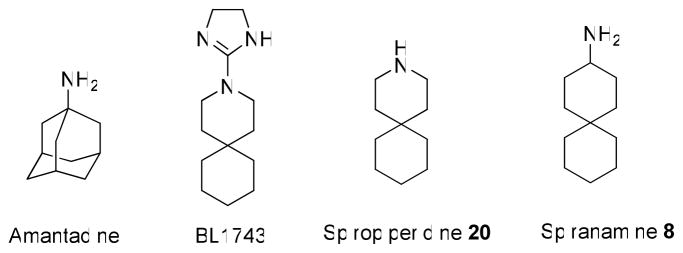
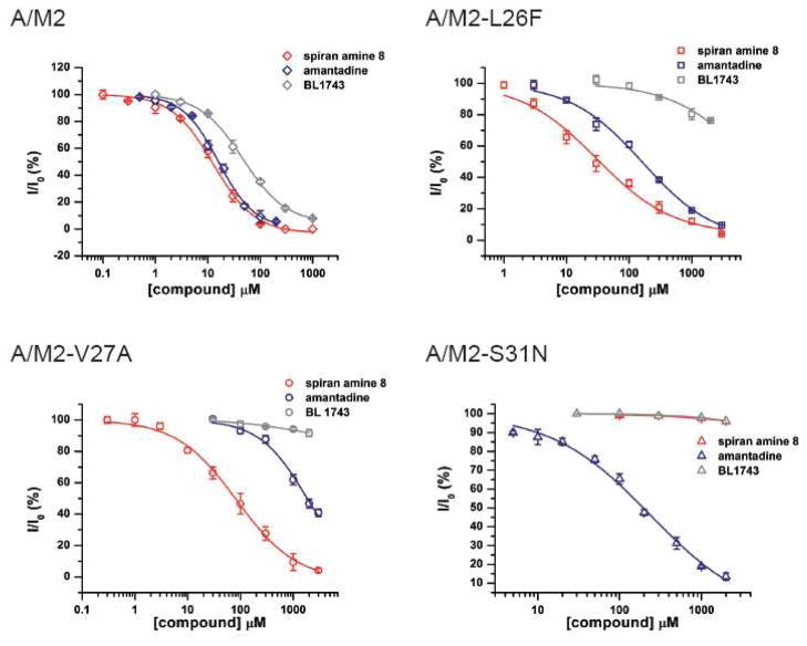
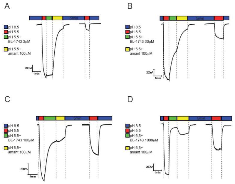
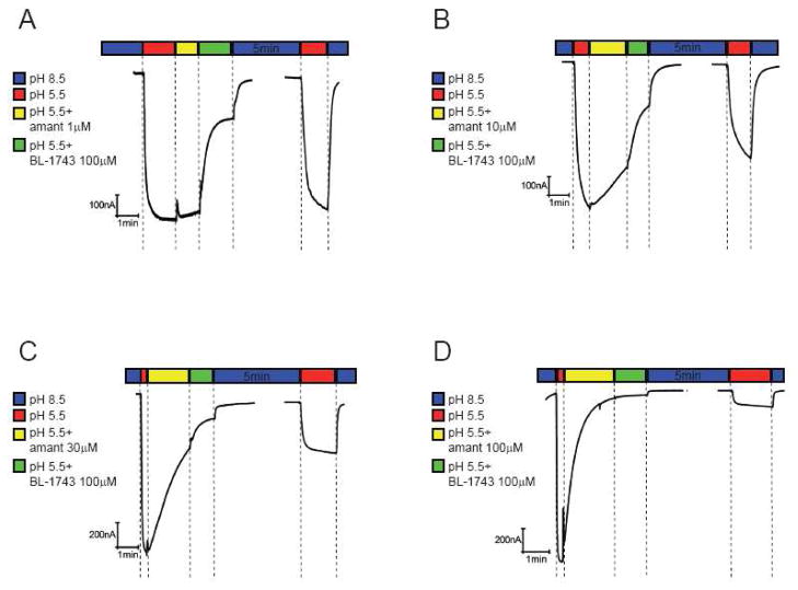
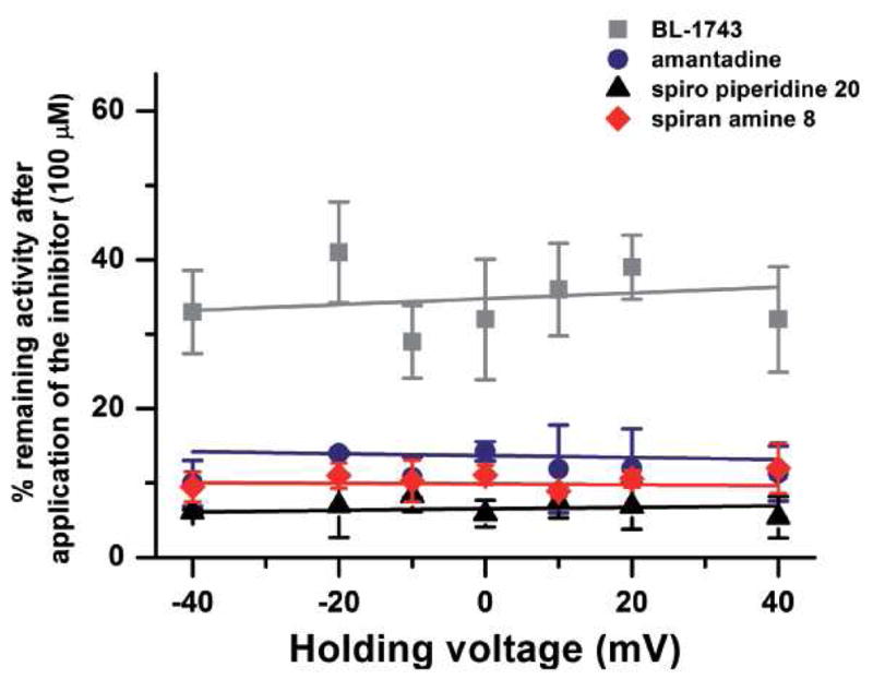
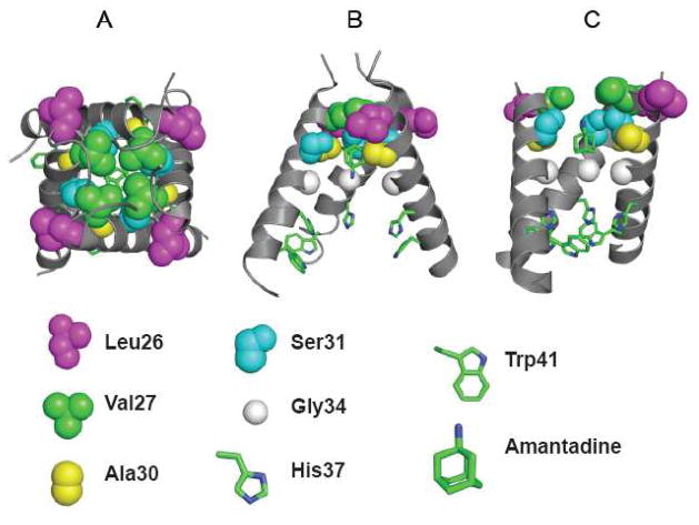
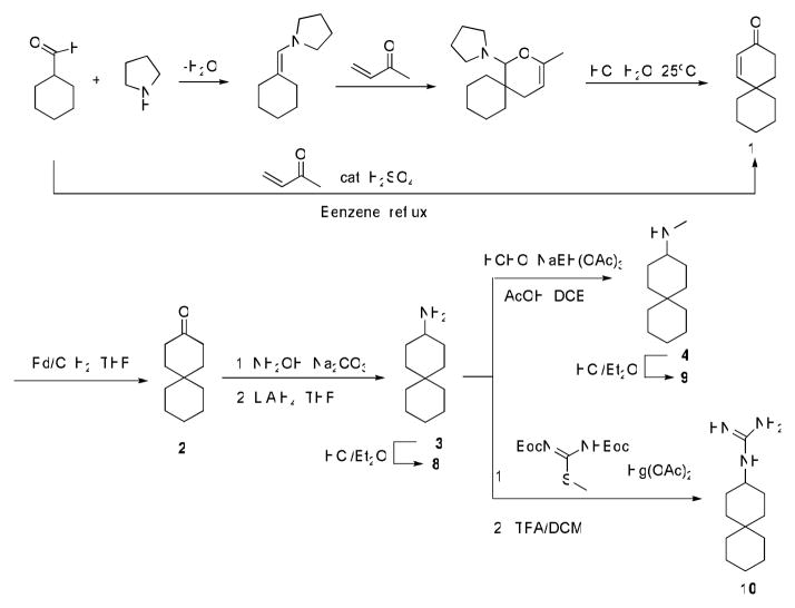
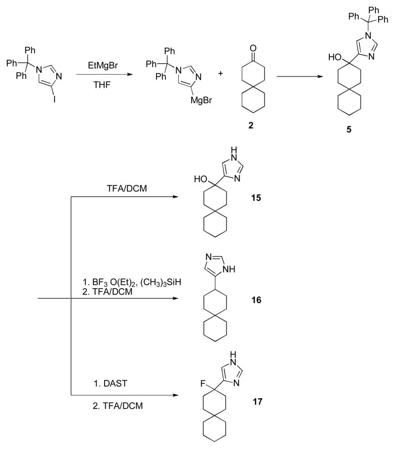
The A/M2 proton channel of influenza A virus is a target for the anti-influenza drugs amantadine and rimantadine, whose effectiveness was diminished by the appearance of naturally occurring point mutants in the A/M2 channel pore, among which the most common are S31N, V27A, and L26F. We have synthesized and characterized the properties of a series of compounds, originally derived from the A/M2 inhibitor BL-1743. A lead compound emerging from these investigations, spiro[5.5]undecan-3-amine, is an effective inhibitor of wild-type A/M2 channels and L26F and V27A mutant ion channels in vitro and also inhibits replication of recombinant mutant viruses bearing these mutations in plaque reduction assays. Differences in the inhibition kinetics between BL-1743, known to bind inside the A/M2 channel pore, and amantadine were exploited to demonstrate competition between these compounds, consistent with the conclusion that amantadine binds inside the channel pore. Inhibition by all of these compounds was shown to be voltage-independent, suggesting that their charged groups are within the N-terminal half of the pore, prior to the selectivity filter that defines the region over which the transmembrane potential occurs. These findings not only help to define the location and mechanism of binding of M2 channel-blocking drugs but also demonstrate the feasibility of discovering new inhibitors that target this binding site in a number of amantadine-resistant mutants.


Figures

Figure 1
Figure 1
Chemical structures of A/M2 channel inhibitors.
Figure 2
Figure 2. Inhibition efficiency of amantadine, BL-1743 and spiran amine 8 on wt A/M2 channels and A/M2 L26F, V27A and S31N mutants
Channel activity was assayed by TEVC for A/M2 channels expressed in Xenopus oocytes. Responses in the presence of various concentrations of an inhibitor (I) were normalized to the current evoked by application of the activating (pH 5.5) solution without inhibitor (I0). The experimental data are the average of three independent experiments. Each point is the mean (±SD) of 5–8 oocytes. IC50 values in μM: A/M2 with amantadine IC50=15.76±1.24; with BL-1743 IC50=46.25±3.56; with spiran amine 8 IC50=12.59±1.11. A/M2-L26F with amantadine IC50=164.46±14.40; with BL-1743 IC50>10 mM; with spiran amine 8 IC50=30.62±8.13. A/M2-V27A with amantadine IC50=1840; with BL-1743 IC50>10 mM; with spiran amine 8 IC50=84.92±13.61. A/M2- S31N with amantadine IC50=237.01±22.14; with BL-1743 IC50>10 mM; with spiran amine 8 IC50>10 mM.
Figure 3
Figure 3. In vivo plaque reduction assay
Wt influenza virus (A/Udorn/72) and influenza virus containing mutations in the M2 TM domain (V27A/L38F) were recovered from cloned DNA and assayed for plaque formation on MDCK cells in the presence or absence of drugs as described in Materials and Methods. A. Effects of amandadine, BL-1743 and compounds 8 and 9 (0.5 and 5 μM) on Udorn plaque formation. B. Effect of BL-1743 (50 and 100 μM) on influenza virus plaque formation. C. Effects of amantadine, BL-1743 and spiran amine 8 (50 μM) on influenza mutant virus V27A/L38F plaque formation. C, upper panel: ~1000 p.f.u./well of mutant virus were used. C, bottom panel: ~100 p.f.u./well of mutant virus were used. Plaque count: no drug – 99 plaques/well; amantadine 50μM – 82 plaques/well; BL-1743 50 μM – 86 plaques/well; spiran amine 8 50 μM – 37 plaques/well.
Figure 4
Figure 4. Inhibition of A/M2 channel activity by saturating concentrations of BL-1743, amantadine and spiran amine
8. A–C; Representative traces of A/M2 channel activity inhibited by either BL-1743 (1000 μM, A), amantadine (100 μM, B), or spiran amine 8 (100 μM, C). A/M2 channel activity was induced by application of the activating solution (pH 5.5, red horizontal bar above the trace). The currents were inhibited by the application of the activating solution containing either BL-1743 (green horizontal bar, A), amantadine (yellow horizontal bar, B), or spiran amine 8 (orange horizontal bar, C) for 60–90 sec. After the maximal inhibition was achieved, oocytes were superfused with the non-activating solution (pH 8.5, second blue horizontal bar) for 5 min to allow current recovery and the channel activity was assayed again by the application of the activating solution (second red horizontal bar). Note that recovery was complete only for BL-1743.
Figure 5
Figure 5. Inhibition of A/M2 channel activity by consecutive application of BL-1743 and amantadine
A–D; Representative traces of the A/M2 channel activity modulated by consecutive application of BL- 1743 and amantadine. A/M2 channel activity was induced by the application of the activating solution (pH 5.5, red horizontal bar above the trace). The currents were first inhibited by the application of the activating solution (pH 5.5) containing various concentrations of BL-1743 (3–1000 μM, green horizontal bar) for 60 sec. After maximal inhibition of the channel activity was achieved, amantadine (100 μM) was substituted for BL-1743 (3–1000 μM) in the activating solution for 90 sec (yellow horizontal bar). The inhibitors were washed out for 5 min by the non-activating solution (pH 8.5, second blue horizontal bar) and the pH-induced activity of A/M2 channels was measured again (second red horizontal bar). Note the biphasic current upon application of amantadine during washout of a saturating concentration of BL-1743 (during yellow bar in D).
Figure 6
Figure 6. Inhibition of A/M2 channel activity by consecutive application of amantadine and BL-1743
A–D, Representative traces of the A/M2 channel activity modulated by consecutive application of amantadine and BL-1743. A/M2 channel activity was induced by the application of the activating solution (pH 5.5, red horizontal bars above the traces). The currents were first inhibited by the application of the activating solution (pH 5.5) containing various concentrations of amantadine (1–100 μM, yellow horizontal bar) for 60 sec. After the maximal inhibition of the channel activity was achieved, BL-1743 (100 μM) was substituted for amantadine (1–100 μM) in the activating for 90 sec (green horizontal bar). The inhibitors were washed out for 5min by the non-activating solution (pH 8.5, second blue horizontal bar) and the pH-induced activity of A/M2 channels was measured again (second red horizontal bar). Note that in no instance was a biphasic current observed during application of BL- 1743 as amantadine was washed-out (yellow bars).
Figure 7
Figure 7. Voltage dependence of A/M2 inhibition by amantadine, BL-1743, spiran amine
8 and spiro piperidine 20. Inhibition of A/M2 channel activity by amantadine, BL-1743, spiran amine 8 and spiro piperidine 20 was assayed at various holding voltages (from −40mV to +40mV). The mean (±SD) channel activity remaining after the maximal inhibition by each of the inhibitors (100 μM) was achieved was plotted as a function of the holding voltage. The experimental data are the average of three independent experiments. Each point is a mean (±SD) of 6–10 oocytes.
Figure 8
Figure 8. Structure of amantadine complexes with the transmembrane domain of A/M2 protein
Panel A and B illustrates the crystal structure of the channel (pdb code 3K9J); viewed from the outside of the virus (A) and in a side-on view with on helix removed for clarity (B). Panel C shows a lower resolution model (2KAD) obtained from an analysis solid-state NMR angular and distance restraints. The identities of critical residues are shown below, showing the C-alpha and sidechain atoms.
Scheme 1
Scheme 1
Synthesis of spiran amine 8, 9 and guanidine 10.
Scheme 2
Scheme 2
Synthesis of spiran triazole 11 and spiran amine 12, 13 and 14 with extended linkers.
Scheme 3
Scheme 3
Synthesis of spiran with imidazole head group 15, 16, 17.
Scheme 4
Scheme 4
Synthesis of spiran 18, 19.

Similar articles

Cited by

References

    1. Cox NJ, Subbarao K. Influenza. Lancet. 1999;354:1277–1282. - PubMed
    1. Lagoja IM, De Clercq E. Anti-influenza virus agents: synthesis and mode of action. Med Res Rev. 2008;28:1–38. - PMC - PubMed
    1. Deyde VM, Xu X, Bright RA, Shaw M, Smith CB, Zhang Y, Shu Y, Gubareva LV, Cox NJ, Klimov AI. Surveillance of resistance to adamantanes among influenza A(H3N2) and A(H1N1) viruses isolated worldwide. J Infect Dis. 2007;196:249–257. - PubMed
    1. Hay AJ, Wolstenholme AJ, Skehel JJ, Smith MH. The molecular basis of the specific anti-influenza action of amantadine. EMBO J. 1985;4:3021–3024. - PMC - PubMed
    1. Garten RJ, Davis CT, Russell CA, Shu B, Lindstrom S, Balish A, Sessions WM, Xu X, Skepner E, Deyde V, Okomo-Adhiambo M, Gubareva L, Barnes J, Smith CB, Emery SL, Hillman MJ, Rivailler P, Smagala J, de Graaf M, Burke DF, Fouchier RA, Pappas C, Alpuche-Aranda CM, Lopez-Gatell H, Olivera H, Lopez I, Myers CA, Faix D, Blair PJ, Yu C, Keene KM, Dotson PD, Jr, Boxrud D, Sambol AR, Abid SH, St George K, Bannerman T, Moore AL, Stringer DJ, Blevins P, Demmler- Harrison GJ, Ginsberg M, Kriner P, Waterman S, Smole S, Guevara HF, Belongia EA, Clark PA, Beatrice ST, Donis R, Katz J, Finelli L, Bridges CB, Shaw M, Jernigan DB, Uyeki TM, Smith DJ, Klimov AI, Cox NJ. Antigenic and Genetic Characteristics of Swine-Origin 2009 A(H1N1) Influenza Viruses Circulating in Humans. Science 2009 - PMC - PubMed

Publication types

MeSH terms