. 2010 Apr;16(4):1339-48.
doi: 10.1089/ten.TEA.2009.0442.

Self-assembly of aligned tissue-engineered annulus fibrosus and intervertebral disc composite via collagen gel contraction

Affiliations

Self-assembly of aligned tissue-engineered annulus fibrosus and intervertebral disc composite via collagen gel contraction

Robby D Bowles et al. Tissue Eng Part A. 2010 Apr.

Abstract

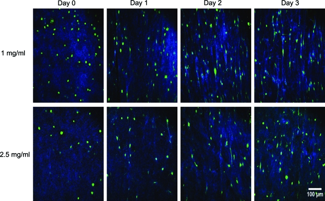
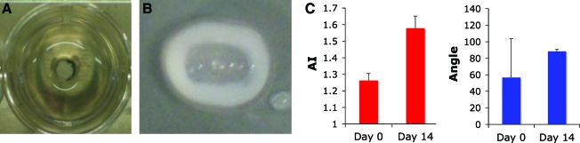
Many cartilaginous tissues such as intervertebral disc (IVD) display a heterogeneous collagen microstructure that results in mechanical anisotropy. These structures are responsible for mechanical function of the tissue and regulate cellular interactions and metabolic responses of cells embedded within these tissues. Using collagen gels seeded with ovine annulus fibrosus cells, constructs of varying structure and heterogeneity were created to mimic the circumferential alignment of the IVD. Alignment was induced within gels by contracting annular gels around an inner boundary using both a polyethylene center and alginate center to create a composite engineered IVD. Collagen alignment and heterogeneity were measured using second harmonic generation microscopy. Decreasing initial collagen density from 2.5 mg/mL to 1 mg/mL produced greater contraction of constructs, resulting in gels that were 55% and 6.2% of the original area after culture, respectively. As a result, more alignment occurred in annular-shaped 1 mg/mL gels compared with 2.5 mg/mL gels (p < 0.05). This alignment was also produced in a composite-engineered IVD with alginate nucleus pulposus. The resulting collagen alignment could promote further aligned collagen development necessary for the creation of a mechanically functional tissue-engineered IVD.


Figures

FIG. 1.
FIG. 1.
Overview of gel culture and imaging methods. Annulus and disk gels were contracted over 3 days before being segmented for imaging. Images were obtained from the outer (o), middle (m), and inner (i) regions of each gel, as defined here. Coordinates were defined in reference to the imaged gel segments. Color images available online at www.liebertonline.com/ten.
FIG. 2.
FIG. 2.
Collagen fiber alignment quantification. (A) SHG microscopy image. (B) Fourier transform of image (contrast adjusted). Fourier amplitude components (FFT image intensities) were summed up along angles at 5° increments from 0 to 180° and represented as an arrow and θ. (C) Summation of amplitudes resulted in a histogram of the image intensities along each 5° increment; from this histogram the mode was calculated, and the alignment index (AI) was calculated according to Equation 1. SHG, second harmonic generation; FFT, fast fourier transform.
FIG. 3.
FIG. 3.
Contraction of disk and annular gels represented as a percentage of the constructs original surface area. Data presented as means ± standard deviations for n = 7 (*p < 0.05).
FIG. 4.
FIG. 4.
SHG-TPEF images (A) from inside region during contraction of 1 and 2.5 mg/mL collagen disk constructs over 3 days, and (B) magnified image showing aligned fibers between cells on day 3 of contraction in 2.5 mg/mL collagen disks (blue, collagen; green, cell). TPEF, two-photon excited fluorescence. Color images available online at www.liebertonline.com/ten.
FIG. 5.
FIG. 5.
SHG-TPEF images from inside region during contraction of 1 and 2.5 mg/mL collagen annular constructs over 3 days. Color images available online at www.liebertonline.com/ten.
FIG. 6.
FIG. 6.
SHG alignment data for collagen annular gels with (A) AI broken down by day and gel concentration (n = 21), (B) AI further broken down by region of gel (n = 7) (O, outside; M, middle; I, inside), and mode angle broken down by day, concentration, and region of gel for (C) 1 mg/mL (n = 4) and (D) 2.5 mg/mL gels (n = 7). Data presented as means and standard deviations (#p < 0.05 compared with day 0; *p < 0.05 for indicated groups). Color images available online at www.liebertonline.com/ten.
FIG. 7.
FIG. 7.
Hematoxylin and eosin staining and TPEF cellular imaging of 1 mg/mL disk gels and annular gels at day 0 and 3 of contraction from inside region of gel.
FIG. 8.
FIG. 8.
(A) Composite disc before contraction with alginate NP in center of well and collagen solution poured around alginate NP. (B) Composite disc after 2 weeks of culture with collagen gel contracted around alginate NP forming tissue-engineered composite intervertebral disc. (C) SHG alignment data measured across entirety of contracted collagen gel thickness indicating high degree of collagen alignment in circumferential direction at day 14. NP, nucleus pulposus. Color images available online at www.liebertonline.com/ten.

References

    1. Ehrlich G.E. Low back pain. Bull World Health Organ. 2003;81:671. - PMC - PubMed
    1. Kelsey J.L. White A.A. 3rd.Epidemiology and impact of low-back pain. Spine. 1980;5:133. - PubMed
    1. Yasuma T. Koh S. Okamura T. Yamauchi Y. Histological changes in aging lumbar intervertebral discs. Their role in protrusions and prolapses. J Bone Joint Surg. 1990;72:220. - PubMed
    1. Kuslich S.D. Ulstrom C.L. Michael C.J. The tissue origin of low back pain and sciatica: a report of pain response to tissue stimulation during operations on the lumbar spine using local anesthesia. Orthop Clin N Am. 1991;22:181. - PubMed
    1. O'Neill C.W. Kurgansky M.E. Derby R. Ryan D.P. Disc stimulation and patterns of referred pain. Spine. 2002;27:2776. - PubMed

Publication types