. 2009;11(6):R82.
doi: 10.1186/bcr2449. Epub 2009 Nov 11.

Dynamic regulation of CD24 and the invasive, CD44posCD24neg phenotype in breast cancer cell lines

Affiliations

Dynamic regulation of CD24 and the invasive, CD44posCD24neg phenotype in breast cancer cell lines

Matthew J Meyer et al. Breast Cancer Res. 2009.

Abstract

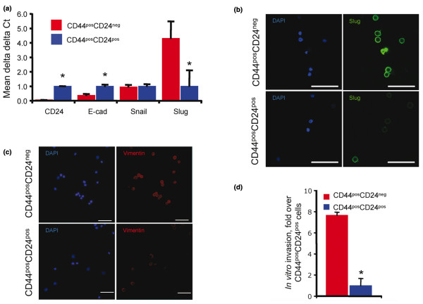
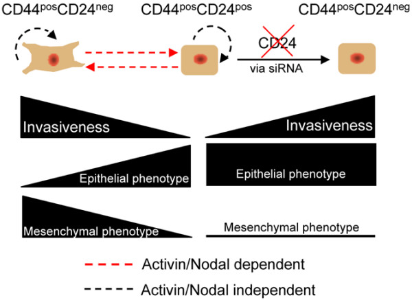
Introduction: The invasive, mesenchymal phenotype of CD44posCD24neg breast cancer cells has made them a promising target for eliminating the metastatic capacity of primary tumors. It has been previously demonstrated that CD44neg/lowCD24pos breast cancer cells lack the ability to give rise to their invasive CD44posCD24neg counterpart. Here we demonstrate that noninvasive, epithelial-like CD44posCD24pos cells readily give rise to invasive, mesenchymal CD44posCD24neg progeny in vivo and in vitro. This interconversion was found to be dependent upon Activin/Nodal signaling.

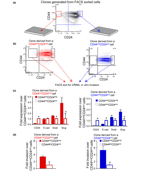
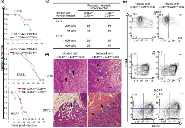
Methods: Breast cancer cell lines were sorted into CD44posCD24pos and CD44posCD24neg populations to evaluate their progeny for the expression of CD44, CD24, and markers of a mesenchymal phenotype. The populations, separated by fluorescence activated cell sorting (FACS) were injected into immunocompromised mice to evaluate their tumorigenicity and invasiveness of the resulting xenografts.

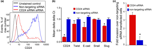
Results: CD24 expression was dynamically regulated in vitro in all evaluated breast cancer cell lines. Furthermore, a single noninvasive, epithelial-like CD44posCD24pos cell had the ability to give rise to invasive, mesenchymal CD44posCD24neg progeny. Importantly, this interconversion occurred in vivo as CD44posCD24pos cells gave rise to xenografts with locally invasive borders as seen in xenografts initiated with CD44posCD24neg cells. Lastly, the ability of CD44posCD24pos cells to give rise to mesenchymal progeny, and vice versa, was blocked upon ablation of Activin/Nodal signaling.

Conclusions: Our data demonstrate that the invasive, mesenchymal CD44posCD24neg phenotype is under dynamic control in breast cancer cell lines both in vitro and in vivo. Furthermore, our observations suggest that therapies targeting CD44posCD24neg tumor cells may have limited success in preventing primary tumor metastasis unless Activin/Nodal signaling is arrested.


Figures

Figure 1
Figure 1
CD24 expression is dynamically regulated in breast cancer cell lines. (a) CD44posCD24neg and CD44posCD24pos cells were sorted from Ca1a, SUM159, MCF7, or MDA MB 231 breast cancer cell lines and expanded in vitro. The CD44, CD24 expression profile of the sorted cells was assessed via flow cytometry after two passages. (b) CD24 promoter CpG islands as predicted by MethPrimer (top panel). A 366 bp region was queried by bisulfate sequencing analysis (BS1, -422 to -788 relative to transcriptional start site). Bisulfite sequencing analysis of CD24 promoter in CD44posCD24pos and CD44posCD24neg parental Ca1a cells. Methylated CG (filled circles) and unmethylated CG (open circles) are presented (middle panel). Percentages of methylation of each CpG in the CD24 promoter from region queried (bottom panel). (c) CD24 mRNA stability assessed in Actinomycin treated, sorted cells. CD44posCD24pos (blue line) and CD44posCD24neg (red line) differences are presented as delta delta Ct means +/- standard deviations around the mean.
Figure 2
Figure 2
CD44posCD24neg cells possess an invasive, mesenchymal phenotype. (a) Total RNA was isolated from CD44posCD24neg and CD44posCD24pos cells sorted from parental Ca1a cells and transcript abundance was evaluated via realtime RT-PCR. CD44posCD24neg (red bars) and CD44posCD24pos (blue bars) differences are presented using the delta delta Ct method. Graphs represent means and associated standard errors of three independent experiments. * indicates P < 0.05. (b, c) Immunofluorescent staining of Slug (green) (b), vimentin (red) (c), and DAPI (blue) in cytospin preparations derived from parental Ca1a cells sorted into CD44posCD24neg and CD44posCD24pos populations. (d) Invasion through Matrigel by sorted parental Ca1a CD44posCD24neg (red bar) and CD44posCD24pos (blue bar) cells. Invaded cells were counted 48 hr following seeding. Graphs represent averages and associated standard errors of experiments performed in triplicate. * indicates P < 0.05.
Figure 3
Figure 3
Clones generated from a single CD44posCD24pos or CD44posCD24neg cell possess molecular and functional heterogeneity similar to that of parental line. (a) Clones were generated by sorting single Ca1a CD44posCD24pos or CD44posCD24neg cells into individual wells of 96-well plates. Only wells confirmed to contain a single cell were expanded. (b) CD44/CD24 expression of representative clones derived from a single CD44posCD24neg or CD44posCD24pos cell. From the experiment described in (a), seven clones were generated from sorted CD44posCD24pos cells and five clones were generated from CD44posCD24neg cells. The CD44/CD24 profiles of representative clones are presented. (c) Total RNA was isolated from isogenic CD44posCD24neg and CD44posCD24pos cells sorted from the clones described in (a, b) and transcript abundance was evaluated via realtime RT-PCR. Data generated with cells sorted from a clone derived from a single CD44posCD24neg cell are presented in red. Data generated with cells sorted from a clone derived from a single CD44posCD24pos cell are presented in blue. CD44posCD24neg (closed bars) and CD44posCD24pos (open bars) differences are presented using the delta delta Ct method. Graphs represent means and associated standard errors of three independent experiments. * indicates P < 0.05. (d) Invasion through Matrigel by isogenic CD44posCD24neg and CD44posCD24poscells freshly sorted from the clones described in (a, b, c). 48 hr following seeding, invaded cells were counted. Graphs represent averages and associated standard errors of experiments performed in triplicate. Data generated with cells sorted from a clone derived from a single CD44posCD24neg cell are presented in red. Data generated with cells sorted from clone derived from a single CD44posCD24pos cell are presented in blue. * indicates P < 0.05.
Figure 4
Figure 4
Xenografts derived from CD44posCD24pos cells are locally invasive and contain CD44posCD24neg progeny. (a) Ca1a (top), ZR75.1 (middle), and MCF7 (bottom) breast cancer cells were FACS sorted based on their CD44, CD24 expression and injected into the abdominal mammary fat pad of nu/nu mice in 50 μl of a 25% Matrigel suspension. Latency of tumor formation following orthotopic injection of sorted CD44posCD24neg/dim (red line) or CD44posCD24pos (blue line) breast cancer cells is presented. Five mice per population were injected and palpated weekly. (b) Limiting dilution of tumor initiating cells in sorted ZR75.1 and Ca1a cells. (c) Xenografts established with either CD44posCD24neg or CD44posCD24pos cells were dissociated and evaluated for CD44/C24 expression. Host cells were excluded with mouse anti-CD45, and -H-2Kd antibodies as described in the Methods section. Representative FACS profiles of xenografts initiated with sorted Ca1a, ZR75.1, and MCF7 cells are presented. (d) Local invasion of xenografts established by injecting CD44posCD24neg or CD44posCD24pos sorted Ca1a or ZR75.1 cells into the abdominal mammary fat pad of nude mice. Representative H&E stained sections of the resulting xenografts are presented. Black arrows indicate host tissue. Light blue arrows indicate invading tumor cells.
Figure 5
Figure 5
Role of Activin/Nodal signaling in the generation of molecular heterogeneity. Ca1a cells were sorted into CD44posCD24neg and CD44posCD24pos populations. Cytospin prepared cells were stained for vimentin immediately following sorting (left two panels, bar represents 200 μm). Sorted cells were allowed to expand for 96 hr in the presence of vehicle (0.1% DMSO, middle two panels) or 10 μM SB-431542 (right two panels). Immunofluorescent staining of vimentin (green) and DAPI (blue) is presented. In the absence of drug, CD44posCD24poscells yield mesenchymal, vimentin positive progeny. Inhibition of Activin/Nodal signaling prevents this interconverstion.
Figure 6
Figure 6
Depletion of CD24 caused increased invasiveness without yielding a mesenchymal phenotype. (a) Ca1a cells were transfected with non-targeting or CD24 siRNA. Histogram illustrates CD24-PE fluorescence intensity of unstained cells (black line) and cells transfected with CD24 siRNA (red line) or non-targeting siRNA (blue line) as assessed by flow cytometric analysis. (b) Total RNA was isolated from cells following transfection with non-targeting or CD24-targeting siRNA. Transcript abundance was evaluated via realtime RT-PCR. Non-targeting siRNA transfected cell (blue bars) and CD24-targeting siRNA transfected cell (red bars) differences are presented using the delta delta Ct method. Graphs represent means and associated standard errors of experiments performed in triplicate. * indicates P < 0.05. (c), 24 hr following transfection with either non-targeting or CD24 siRNA, Ca1a cells were trypsinized, counted, and seeded to Matrigel invasion chambers in triplicate. Invaded cells were counted 48 hr later. Graph represents mean fold change in invaded cells associated with CD24 siRNA transfection and associated standard errors. * indicates P < 0.05.
Figure 7
Figure 7
Dynamic regulation of CD24 and the invasive CD44posCD24neg phenotype. Schematic representation of the plasticity of the CD44posCD24neg, invasive/mesenchymal phenotype. Inhibition of Activin/Nodal signaling blocks the ability of CD44posCD24pos cells to give rise to invasive, mesenchymal progeny and CD44posCD24neg cells are prevented from giving rise to non-invasive, epithelial progeny. Depletion of CD24 via siRNA causes increased invasiveness without yielding a mesenchymal phenotype.

References

    1. Kristiansen G, Sammar M, Altevogt P. Tumour biological aspects of CD24, a mucin-like adhesion molecule. J Mol Histol. 2004;35:255–262. doi: 10.1023/B:HIJO.0000032357.16261.c5. - DOI - PubMed
    1. Friederichs J, Zeller Y, Hafezi-Moghadam A, Grone HJ, Ley K, Altevogt P. The CD24/P-selectin binding pathway initiates lung arrest of human A125 adenocarcinoma cells. Cancer Res. 2000;60:6714–6722. - PubMed
    1. Fischer GF, Majdic O, Gadd S, Knapp W. Signal transduction in lymphocytic and myeloid cells via CD24, a new member of phosphoinositol-anchored membrane molecules. J Immunol. 1990;144:638–641. - PubMed
    1. Shipitsin M, Campbell LL, Argani P, Weremowicz S, Bloushtain-Qimron N, Yao J, Nikolskaya T, Serebryiskaya T, Beroukhim R, Hu M, Halushka MK, Sukumar S, Parker LM, Anderson KS, Harris LN, Garber JE, Richardson AL, Schnitt SJ, Nikolsky Y, Gelman RS, Polyak K. Molecular definition of breast tumor heterogeneity. Cancer Cell. 2007;11:259–273. doi: 10.1016/j.ccr.2007.01.013. - DOI - PubMed
    1. Villadsen R. In search of a stem cell hierarchy in the human breast and its relevance to breast cancer evolution. APMIS. 2005;113:903–921. doi: 10.1111/j.1600-0463.2005.apm_344.x. - DOI - PubMed

Publication types

MeSH terms