. 2010 Jan 5;396(1):118-24.
doi: 10.1016/j.virol.2009.10.018. Epub 2009 Nov 10.

Destabilization of Rb by human papillomavirus E7 is cell cycle dependent: E2-25K is involved in the proteolysis

Affiliations

Destabilization of Rb by human papillomavirus E7 is cell cycle dependent: E2-25K is involved in the proteolysis

Kwang-Jin Oh et al. Virology. .

Abstract

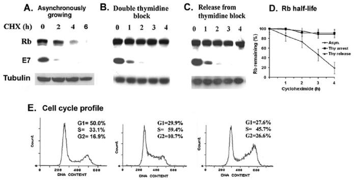
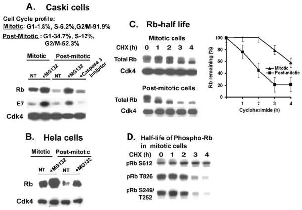
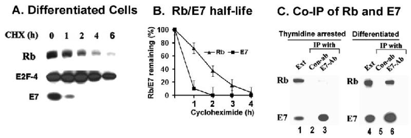
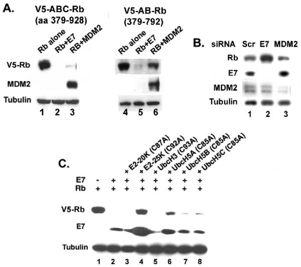
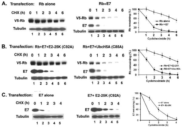
The HPV oncoprotein E7 promotes proteasomal degradation of the tumor suppressor protein Rb. In this study, we analyzed the regulation of E7-induced Rb proteolysis in HPV-containing Caski cervical cancer cells. We show that the Rb proteolysis is cell cycle dependent; in S phase Rb is stable while in post-mitotic early G1 phase cells and in differentiated cells, Rb is unstable. Similarly, the in vivo Rb/E7 interaction is not detected in S-phase cells, but is readily detected in differentiating Caski cells. The ubiquitinating enzymes involved in Rb proteolysis have not been identified. We find that the E3 ligase MDM2 is not involved in the Rb proteolysis in Caski cells. An in vivo analysis using multiple catalytic site mutant dominant negative E2 enzymes show that the C92A E2-25K most effectively blocks E7-induced Rb proteolysis. Taken together, these results show that E7 induces Rb proteolysis in growth-arrested cells and E2-25K is involved in the proteolysis.


Figures

Figure 1
Figure 1
Rb proteolysis is blocked in the S-phase of Caski cells. Caski cells were arrested in S-phase by double thymidine block as described in the materials and methods. Asynchronously growing cells (panel A), thymidine arrested cells (panel B), and thymidine-released cells (panel C) were treated with cycloheximide (25ug/ml) for the indicated time. Cell lysates were analyzed for Rb, E7, and α-tubulin (loading control) using the Western blot assay. (D). A quantification of the Rb band intensity was plotted against the time after the cycloheximide addition. The average of two independent experiments is shown. (E) Cell cycle profiles of asynchronous, thymidine arrested and thymidine released Caski cells are shown.
Figure 2
Figure 2
(A-B) Proteasomal degradation of Rb is enhanced in post-mitotic cells. (A) Caski cells were arrested at the M phase by growing in nocodazole (100ng/ml) containing medium for 20h, and the mitotic cells were then grown in fresh medium for 4h to get the G1-enriched cells. MG132 (10μM) and Caspase 3 inhibitor III (100μM) was added for the last 4h prior to harvesting. Cell cycle profiles of mitotic and post-mitotic Caski cells are shown. Cell lysates were analyzed for Rb, E7, and Cdk4 (loading control). (B) Mitotic and post-mitotic HeLa cells were treated with MG132 (10μM) for 4h, and the cell lysates were analyzed for Rb and Cdk4 (loading control). (C) Lower half-life of Rb in post-mitotic cells during M to G1 progression. Cycloheximide (25ug/ml) was added to the mitotic and post mitotic cells, and cells were harvested every hour for up to 4h. Left panel: Cell lysates from mitotic cells, and post-mitotic cells were analyzed for Rb and Cdk4 (loading control). Right panel: A quantification of total Rb band intensities in mitotic and post-mitotic cells was plotted against time after the cycloheximide addition. An average of two independent experiments is shown. (D) Different half-lives of phospho-Rb in Caski cells. Mitotic cell lysates were analyzed for the phospho-Rb species, pRbS612, pRbT826, and pRb S249/T252 using western blot assay.
Figure 3
Figure 3
(A) Rb proteolysis is enhanced in differentiated cells. Caski cells were differentiated by culturing in 1.6%methyl cellulose containing medium for 14h. Cycloheximide (50ug/ml) was added, and the cell lysates (150ug) were analyzed for Rb, E7, and E2F-4 (loading control). (B) A quantification of Rb and E7 band intensities was plotted against time after cycloheximide addition in differentiated cells. (C) The Rb-E7 interaction is cell cycle dependent. Cell lysates (1mg) from differentiated Caski cells (right panel), and thymidine-arrested Caski cells (left panel) were immunoprecipitated with either control antibody or the polyclonal E7-antibody. The immunoprecipitated proteins were analyzed for Rb and E7 using the monoclonal antibodies.
Figure 4
Figure 4
(A-B) E7-induced Rb proteolysis does not involve MDM2. (A) C-terminal sequences of Rb are critical for proteolysis by MDM2 not E7. C33A cells were transfected with plasmids expressing V5-epitope tagged ABC-Rb (aa379-928); V5-epitope tagged AB-Rb (379-792) with HPV16 E7 or MDM2. The cell lysates were analyzed for Rb, MDM2 and tubulin (loading control). (B) MDM2 siRNA does not increase the steady state level of Rb in Caski cells. Caski cells were transfected with control siRNA, E7 siRNA, and MDM2 siRNA. After 48h, the cells were harvested and the level of Rb, MDM2, E7, and tubulin (loading control) were analyzed. (C) The catalytic site mutant C92A E2-25K restores the level of Rb in presence of E7. C33A cells were transfected with plasmids expressing V5-ABC-Rb, HPV16 E7, and the active site Cys-Ala mutants of different E2 enzymes as indicated. The cell lysates were analyzed for V5-Rb, E7, and tubulin (loading control).
Figure 5
Figure 5
(A-B). The catalytic site mutant C92A E2-25K inhibits E7-mediated proteolysis of Rb. C33A cells were transfected with plasmids expressing V5-ABC-Rb alone, V5-ABC-Rb and HPV16 E7 (panel A); V5-ABC-Rb, HPV16 E7, C92A E2-25K and V5-ABC-Rb, HPV16 E7, C85A UbcH5A (panel B). 36h after transfection, the cells were incubated with cycloheximide (50ug/ml) for the indicated times. The cell lysates were analyzed for V5-Rb, E7, and tubulin (loading control). (C). C92A E2-25K partially stabilizes HPV16 E7. C33A cells were transfected with plasmids expressing HPV16 E7, and HPV16 E7 and C92A E2-25K, incubated with cycloheximide and analyzed for E7 and tubulin (loading control). Quantitations of the band intensities of Rb and E7 against the time after cycloheximide addition are shown.

References

    1. Berezutskaya E, Morozov A, Raychaudhuri P, Bagchi S. Differential regulation of the pocket domains of the retinoblastoma family proteins by the HPV16E7 oncoprotein. Cell Growth and Differ. 1997;8:1277–1286. - PubMed
    1. Binné U,K, Classon MK, Dick FA, Wei W, Rape M, Kaelin WG, Jr., Näär AM, Dyson NJ. Retinoblastoma protein and anaphase-promoting complex physically interact and functionally cooperate during cell-cycle exit. Nat Cell Biol. 2007;9:225–232. - PubMed
    1. Boyer SN, Wazer DE, Band V. E7 protein of human papillomavirus-16 induces degradation of retinoblastoma protein through the ubiquitin–proteasome pathway. Cancer Res. 1996;56:4620–4624. - PubMed
    1. Cheng S, Schmidt-Grimminger DC, Murant T, Broker TR, Chow LT. Differentiation-dependent up-regulation of the human papillomavirus E7 gene reactivates cellular DNA replication in suprabasal differentiated keratinocytes. Genes Dev. 1995;9:2335–2349. - PubMed
    1. Cobrinik D. Pocket proteins and cell cycle control. Oncogene. 2005;24:2796–2809. - PubMed

Publication types

MeSH terms

Substances