. 2010 Feb;31(6):1235-41.
doi: 10.1016/j.biomaterials.2009.10.052. Epub 2009 Nov 10.

Effects of VEGF temporal and spatial presentation on angiogenesis

Affiliations

Effects of VEGF temporal and spatial presentation on angiogenesis

Eduardo A Silva et al. Biomaterials. 2010 Feb.

Abstract

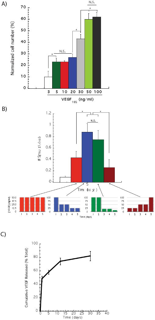
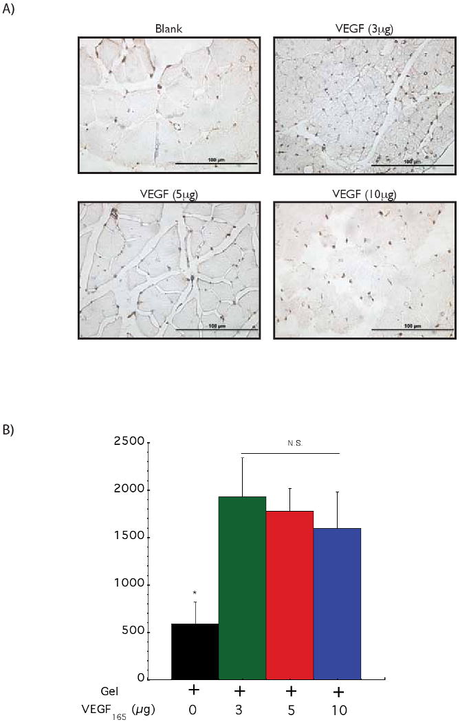
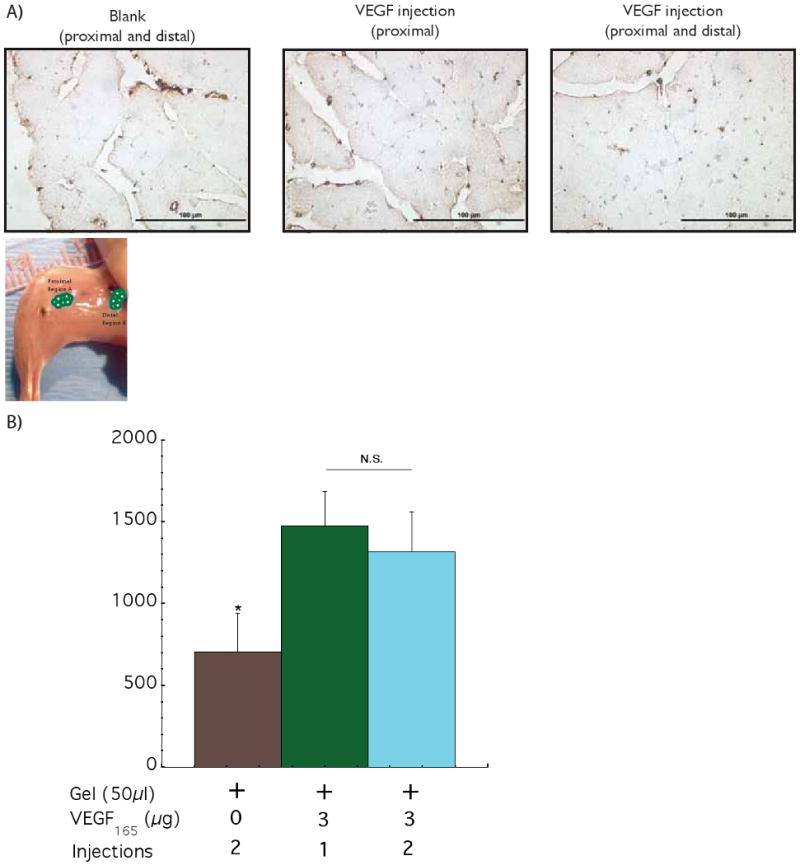
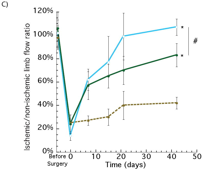
Therapeutic angiogenesis relies on the delivery of angiogenic factors capable of reversing tissue ischemia. Polymeric materials that can provide spatial and temporal over vascular endothelial growth factor (VEGF) presentation provide clear benefit, but the influence of VEGF dose, temporal, and spatial presentation on the resultant angiogenic process are largely unknown. The influence of the temporal profile of VEGF concentration, dose, and the impact of VEGF spatial distribution on angiogenesis in in vitro models of angiogenesis and ischemic murine limbs was analyzed in this study. Importantly, a profile consisting of a high VEGF concentration initially, followed by a decreasing concentration over time was found to yield optimal angiogenic sprouting. A total VEGF dose 0.1 microg/g, when delivered with kinetics found to be optimal in vitro, provided a favorable therapeutic dose in murine hindlimb ischemia model, and distributing this VEGF dose in two spatial locations induces a higher level of vascularization and perfusion than a single location. These findings suggest that material systems capable of controlling and regulating the temporal and spatial presentation of VEGF maybe useful to achieve a robust and potent therapeutic angiogenic effect in vivo.


Figures

Figure 1
Figure 1
Effect of VEGF dose on in vitro endothelial cell proliferation and sprouting. The proliferation of human microvascular endothelial cells (HMVEC-d) was analyzed after 73 hours of culture with distinct VEGF165 concentrations (A). A statistically significant higher endothelial cell proliferation was observed when cells were cultured with 50 ng/ml of VEGF165, as compared to lower VEGF concentrations, but no statistically difference was noted between 50 and 100 ng/ml. Gradually decreasing the VEGF dose induced a greater number of endothelial cells sprouts, as compared to a constant VEGF doses (50 ng/ml day), or a gradual VEGF dose decrease over time (B). The release kinetics of 125I – VEGF165 from gels formed from binary molecular weight alginate partially oxidized was monitored over time (C). In (A) (B) and (C), values represent mean and standard deviation (A, n=6 and B and C, n=4).
Figure 2
Figure 2
Monitoring the effect of VEGF dose in driving vascularization in ApoE-/- ischemic hindlimbs. Representative photomicrographs from CD31 immunostained sections of hindlimb muscle tissues (A). Quantification of blood vessel densities in hindlimb muscle tissues 6 weeks after treatment with a control blank gel (+ 0); a gel with 3 μg of VEGF (+ 3); a gel with 5 μg of VEGF (+ 5) and a gel with 10 μg of VEGF (+ 10) (B). No statistically significant differences were observed between the different VEGF doses. Tissue perfusion of ApoE-/- mice hindlimbs at various time points following treatment with a control blank gel (◇); 3 μg of VEGF delivered from alginate hydrogels (●); 5 μg of VEGF delivered from alginate hydrogels (○) and 10 μg of VEGF delivered from alginate hydrogels (□) (C). Mean values are presented with standard deviations and * indicates statistically significant differences (p<0.05), as compared to control gels. N.S. displays no statistically significant difference between conditions.
Figure 2
Figure 2
Monitoring the effect of VEGF dose in driving vascularization in ApoE-/- ischemic hindlimbs. Representative photomicrographs from CD31 immunostained sections of hindlimb muscle tissues (A). Quantification of blood vessel densities in hindlimb muscle tissues 6 weeks after treatment with a control blank gel (+ 0); a gel with 3 μg of VEGF (+ 3); a gel with 5 μg of VEGF (+ 5) and a gel with 10 μg of VEGF (+ 10) (B). No statistically significant differences were observed between the different VEGF doses. Tissue perfusion of ApoE-/- mice hindlimbs at various time points following treatment with a control blank gel (◇); 3 μg of VEGF delivered from alginate hydrogels (●); 5 μg of VEGF delivered from alginate hydrogels (○) and 10 μg of VEGF delivered from alginate hydrogels (□) (C). Mean values are presented with standard deviations and * indicates statistically significant differences (p<0.05), as compared to control gels. N.S. displays no statistically significant difference between conditions.
Figure 3
Figure 3
Assessing the effect of spatial distribution of VEGF delivery on the ischemic ApoE-/- hindlimbs. Photomicrographs of representative tissue sections from hindlimbs of ApoE-/- mice at postoperative 42 days, immunostained for the endothelial marker CD31 (A). Quantification of blood vessels densities after 6 weeks with 2 injections (proximal and distal) of control blank alginate hydrogel (+ 0 2); 1 injection of alginate loaded with VEGF (proximal) (+ 3 1) and 2 injections of alginate loaded with VEGF (proximal and distal) (+ 3 2) (B). Similar values of blood vessel densities were obtained for the 1 and 2 injections of VEGF. Perfusion profiles of hindlimbs at various experimental time points with 2 injections of control blank gel (●); VEGF delivered from alginate gels (1 injection - proximal) (●) and VEGF delivered from alginate gels (2 injections – proximal and distal) (○) (C). The two injections of VEGF elicited an increase in the regional perfusion, as compared with a single injection (VEGF loaded), even though the total VEGF dose was the same between the two conditions. Mean values are presented with standard deviations, * indicates statistically significant differences (p<0.05), as compared to control gels, and # represents statistically significant differences (p<0.05) between conditions.
Figure 3
Figure 3
Assessing the effect of spatial distribution of VEGF delivery on the ischemic ApoE-/- hindlimbs. Photomicrographs of representative tissue sections from hindlimbs of ApoE-/- mice at postoperative 42 days, immunostained for the endothelial marker CD31 (A). Quantification of blood vessels densities after 6 weeks with 2 injections (proximal and distal) of control blank alginate hydrogel (+ 0 2); 1 injection of alginate loaded with VEGF (proximal) (+ 3 1) and 2 injections of alginate loaded with VEGF (proximal and distal) (+ 3 2) (B). Similar values of blood vessel densities were obtained for the 1 and 2 injections of VEGF. Perfusion profiles of hindlimbs at various experimental time points with 2 injections of control blank gel (●); VEGF delivered from alginate gels (1 injection - proximal) (●) and VEGF delivered from alginate gels (2 injections – proximal and distal) (○) (C). The two injections of VEGF elicited an increase in the regional perfusion, as compared with a single injection (VEGF loaded), even though the total VEGF dose was the same between the two conditions. Mean values are presented with standard deviations, * indicates statistically significant differences (p<0.05), as compared to control gels, and # represents statistically significant differences (p<0.05) between conditions.

References

    1. Mathers CD, Loncar D. Projections of global mortality and burden of disease from 2002 to 2030. PLoS Med. 2006;3:e442. - PMC - PubMed
    1. Abegunde DO, Mathers CD, Adam T, Ortegon M, Strong K. The burden and costs of chronic diseases in low-income and middle-income countries. Lancet. 2007;370:1929–1938. - PubMed
    1. Place ES, Evans ND, Stevens MM. Complexity in biomaterials for tissue engineering. Nat Mater. 2009;8:457–470. - PubMed
    1. Carmeliet P. Angiogenesis in life, disease and medicine. Nature. 2005;438:932–936. - PubMed
    1. Folkman J. Angiogenesis: an organizing principle for drug discovery? Nat Rev Drug Discov. 2007;6:273–286. - PubMed

Publication types

MeSH terms

Substances