Review
. 2009 Sep 15;19(17):R749-61.
doi: 10.1016/j.cub.2009.07.028.

Force and length in the mitotic spindle

Affiliations
Review

Force and length in the mitotic spindle

Sophie Dumont et al. Curr Biol. .

Abstract

The mitotic spindle assembles to a steady-state length at metaphase through the integrated action of molecular mechanisms that generate and respond to mechanical forces. While molecular mechanisms that produce force have been described, our understanding of how they integrate with each other, and with the assembly/disassembly mechanisms that regulate length, is poor. We review current understanding of the basic architecture and dynamics of the metaphase spindle, and some of the elementary force-producing mechanisms. We then discuss models for force integration and spindle length determination. We also emphasize key missing data that notably include absolute values of forces and how they vary as a function of position within the spindle.


Figures
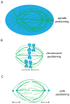
Figure 1
Figure 1
Three steady-states in position are reached during metaphase. Position-dependent forces (black arrows) must help reach the steady-state positions and correct any deviations (fainter colors) from them. A) During symmetrical cell division, the spindle (green) must be positioned at the center of the cell (blue). B) The chromosomes (blue) must be placed in the middle of the two poles. C) The spindle poles (blue) must be positioned a certain distance away from each other (and the chromosomes) to dictate spindle length. The three steady-state positions are critical in determining where the sister chromosomes will travel after anaphase, and thus essential to accurate chromosome segregation.
Figure 2
Figure 2
Microtubule architecture and dynamics in the mitotic metaphase spindle of mammalian cells. A) Architecture of the mammalian mitotic spindle: microtubules (green), sister chromosomes (blue) and kinetochores (red) for attachment of chromosomes to microtubules. B–D) Three classes of microtubules within the spindle, with different minus-end locations (black circles), dynamics (black arrows) and nucleation zones (blue). B) Kinetochore-microtubules continuously slide toward the pole (green arrow), polymerize at the kinetochore and depolymerize at the pole. Kinetochore-microtubules form larges bundles (thicker green line) and have much longer lifetimes than the other two classes of microtubules. C) Nonkinetochore-microtubules are nucleated throughout the spindle, and continuously slide poleward (green arrow) with dynamic plus-ends and unprobed minus-ends. D) Astral microtubules are nucleated at centrosomes, don’t slide, have dynamic plus-ends and fixed minus-ends. Astral microtubules may also overlap with other microtubules (question mark).
Figure 3
Figure 3
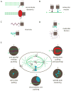
Molecular force generators and their sites of action in the mammalian metaphase spindle. Arrows depict object (square) direction of movement (small arrows) and experienced force (large arrows). A) Microtubules (green) assembling (top) and disassembling (bottom) can push or pull an object, respectively. To couple disassembly to object movement, a connecting element (red ellipse) is required. B) A molecular motor can power object movement toward microtubule plus-ends (purple) or minus-ends (blue). C) An elastic element (spring) can pull objects inward when stretched, or push objects outward when compressed. D) Friction forces oppose movement. They can be generated by bond breakage (top, blue bonds moving up and down) and mesh reorganization (bottom) required for object movement. E) Spindle locations where the above forces operate. Only dominant forces are cartooned. Friction and elasticity likely operate everywhere, but are only drawn at the kinetochore. (1) Anywhere anti-parallel microtubules overlap, microtubule cross-linking motors operate. This site is depicted both near and far from the metaphase plate. Kinesin-5 family members push microtubules apart: this is the best characterized outward force, and is required for bipolarity establishment in most spindles [44, 97]. C. elegans embryonic spindles largely lack nK-MTs and do not require this force [122]. (2) At kinetochores, where microtubules disassemble and pull (and assemble and may push), and where Ndc80 (red arms) is thought to provide microtubule attachment. The elastic centromere (spring) is deformed [76] and friction (double arrow) occurs. Plus- and minus-end motors (e.g. Cenp-e and Dynein) can also operate here, as can microtubule depolymerases (e.g. MCAK and Kif18) and other end binding proteins [13, 26, 75, 123, 124]. (3) At the poles, dynein and/or minus-end kinesins organize and focus minus-ends, presumably by holding on to one microtubule while moving on another [125]. K-MTs depolymerize at poles (depolymerases may be involved [85]), and whether this generates pulling forces has been suggested [37, 84] but not directly measured. (4) On chromosome arms, plus-end-directed chromokinesins (e.g Kid [98]) push microtubules, exerting away-from-the-pole force (polar ejection force [104]). (5) At the cortex, dynein pulls on A-MTs, and may be the main spindle-centering force in mammalian cells [126]. How motor activity is coupled to depolymerization (and polymerization) at the cortex is unclear.
Figure 4
Figure 4
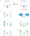
Toward a primitive force map of the mammalian metaphase spindle. Experiments informing on mechanical properties of the spindle. A) Classic tensed k-fibers force map (left), and revised force map proposed herein (right). Red bar represents a possible non-microtubule element under tension. B) Laser ablating one kinetochore results in poleward movement of the sister kinetochore [83]. C) Addition of nocodazole results in reduced tension on the kinetochores [61]. D) Cutting several k-fibers results in bending of the few remaining fibers and shortening of that half-spindle [87]. E) Release of a microtubule depolymerizer drug results in bending of the stable k-fibers and loss of kinetochore tension as the spindle shortens [88]. F) Laser cutting a k-fiber near the kinetochore does not prevent tension generation on that kinetochore, or microtubule sliding (green arrow) [34, 87]. G) A microneedle can move a chromosome across the metaphase plate while the k-fiber stays connected at the pole [31, 32].
Figure 5
Figure 5
Nature of the metaphase spindle length problem. A) For small cells, spindle length scales with cell size, but for larger cells [100] and in extract [101], spindle length reaches an upper limit. B) The spindle is a dynamic structure. Physical perturbations reversibly change the spindle length steady-state: the spindle lengthens upon egg [4] or spindle [2, 8] compression, and shortens when subject to high hydrostatic pressure [9], low temperature [5, 6], or pole-to-pole microneedle compression [3]. Similarly, addition of hexylene glycol [88] or D2O [127] increases spindle length, while colchicine reversibly decreases spindle length [4]. While these physical and chemical perturbations affect total spindle tubulin polymer mass, we do not know whether they affect spindle length by changing microtubule length, growth parameters or numbers in the spindle. Genetic perturbations that affect both spindle assembly and maintenance (e.g. RNAi, depletions) revealed that microtubule destabilizers contribute to spindle shortening [88, 114], and microtubule stabilizers [114] and nucleators [128] contribute to lengthening; the location of destabilizers may be important, and their activities may oppose each other [129]. Chemical and genetic perturbations of motors [37, 44, 114], and kinetochore-microtubule attachment [68], can also affect spindle length and are not included here; their role may be system-dependent. C) Bipolar and monopolar spindles have the same chromosome-to-pole distance [104]. Purple arrows represent the position-dependence of polar ejection forces [109] (powered in part by chromokinesins, see Fig. 3E).
Figure 6
Figure 6
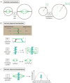
Three classes of models able to provide a stable metaphase spindle length scale. A) Spindle-extrinsic mechanisms. For example, cell size (A1) or availability of a spindle component (e.g. tubulin monomer) (A2) could determine spindle length. B) Spindle-intrinsic physical mechanisms: inward forces could increase with spindle length (left), or outward forces could decrease with spindle length (right). Proposed mechanisms include opposed motors (B1), a slide-and-cluster model (B2, where half-spindle length is proportional to the product of microtubule sliding velocity v and lifetime Δt), and an elastic structural scaffold (B3). C) Spindle-intrinsic chemical mechanisms. For example, a morphogen (grey molecule) gradient (C1) could determine spindle length. Right cartoon represents the morphogen concentration decay away from chromosomes; the dotted lines represent the concentration threshold determining pole position.

Similar articles

Cited by

References

    1. Walczak CE, Heald R. Mechanisms of mitotic spindle assembly and function. International Review of Cytology. 2008;265:111–158. - PubMed
    1. Dumont S, Mitchison TJ. Compression regulates spindle length by a mechanochemical switch at the poles. Current Biology. 2009 Jun 17; [Epub ahead of print] - PMC - PubMed
    1. Hays TS. The force-balance mechanism of chromosome congression. Chapel Hill: University of North Carolina at Chapel Hill; 1985. p. 156. Volume Ph.D.
    1. Inoué S. The effect of colchicine on the microscopic and submicroscopic structure of the mitotic spindle. Experimental Cell Research Supplement. 1952;2:305–318.
    1. Inoué S. Effect of temperature on the birefringence of the mitotic spindle. Biological Bulletin. 1952;103:316.

Publication types

LinkOut - more resources