Review
. 2009 Sep 15;19(17):R762-71.
doi: 10.1016/j.cub.2009.06.053.

The shape of motile cells

Affiliations
Review

The shape of motile cells

Alex Mogilner et al. Curr Biol. .

Abstract

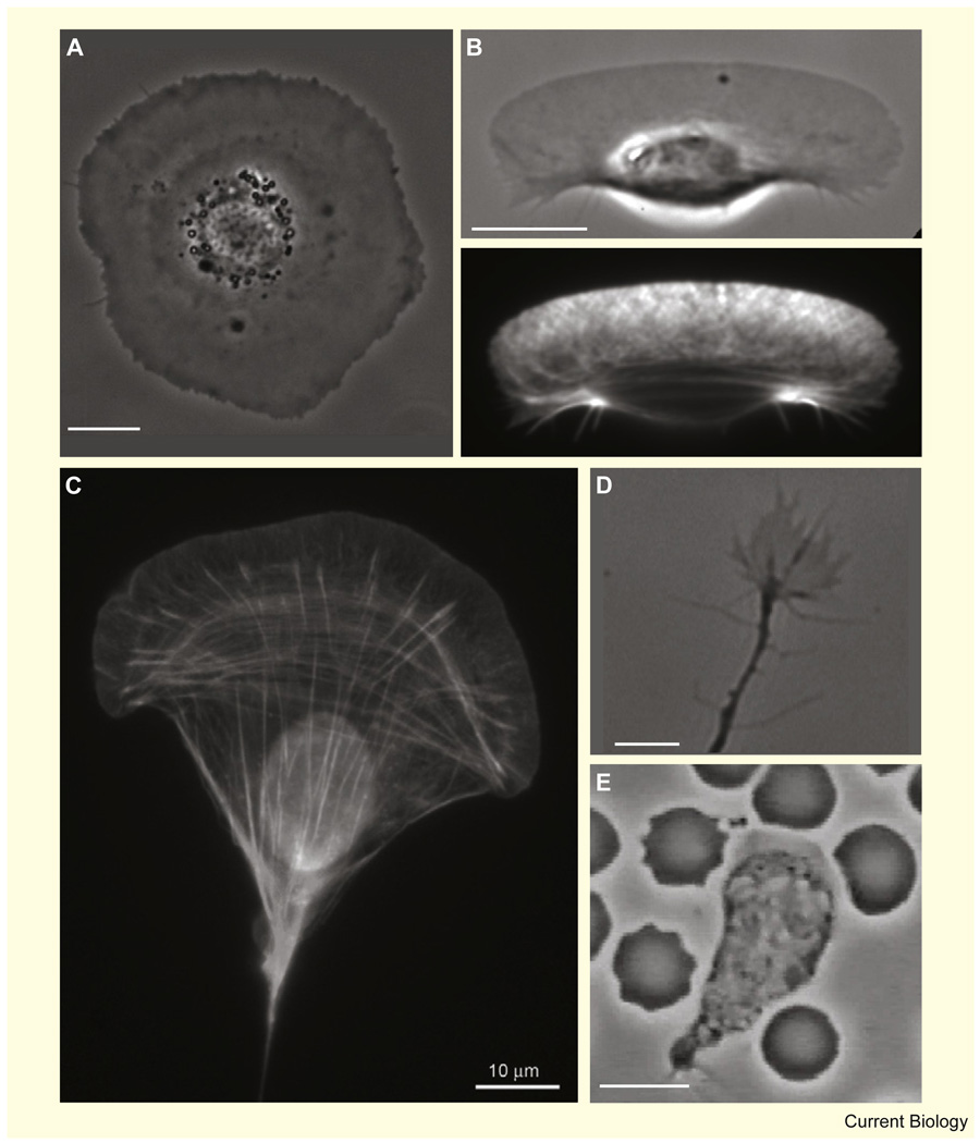
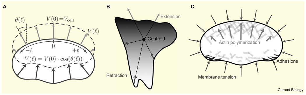
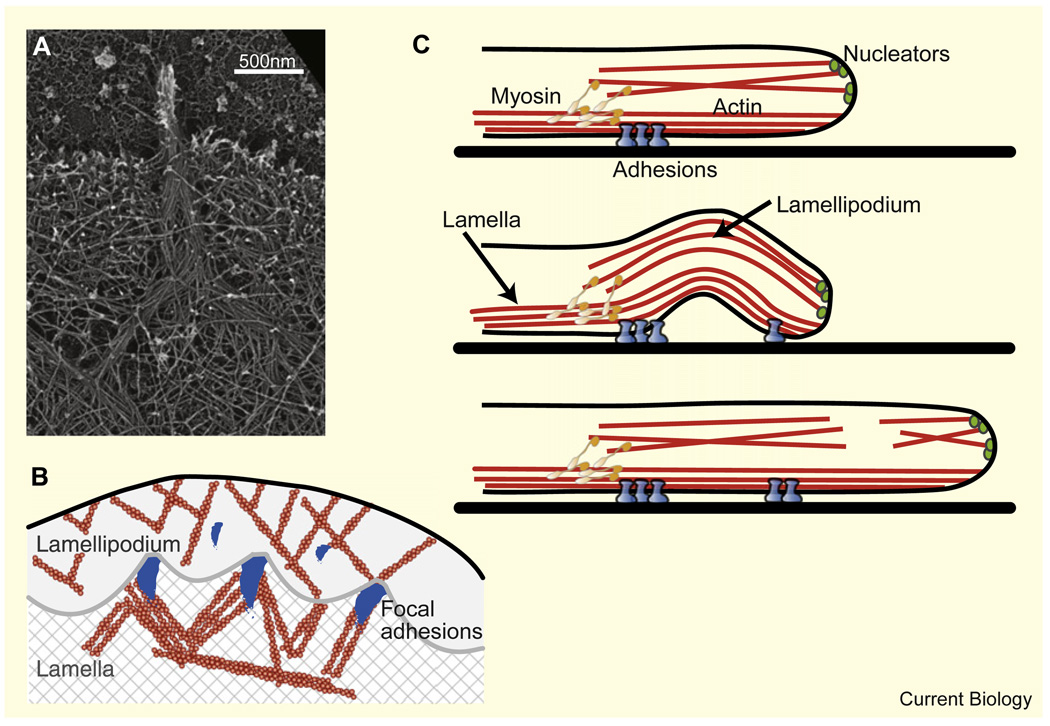
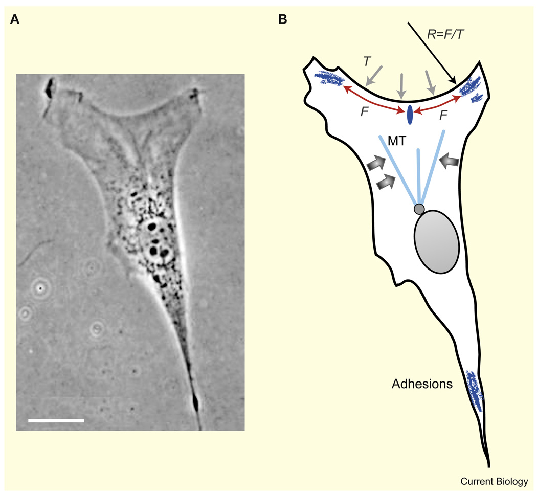
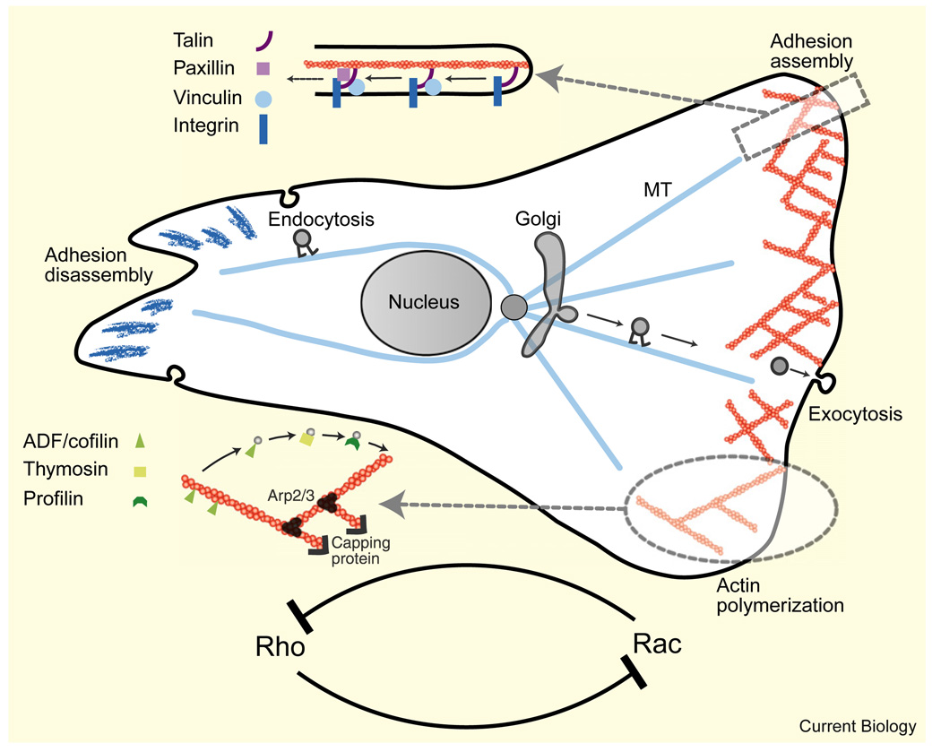
Motile cells - fan-like keratocytes, hand-shaped nerve growth cones, polygonal fibroblasts, to name but a few - come in different shapes and sizes. We discuss the origins of this diversity as well as what shape tells us about the physics and biochemistry underlying cell movement. We start with geometric rules describing cell-edge kinetics that govern cell shape, followed by a discussion of the underlying biophysics; we consider actin treadmilling, actin-myosin contraction, cell-membrane deformations, adhesion, and the complex interactions between these modules, as well as their regulation by microtubules and Rho GTPases. Focusing on several different cell types, including keratocytes and fibroblasts, we discuss how dynamic cell morphology emerges from the interplay between the different motility modules and the environment.


Figures

Figure 1
Figure 1. Diverse shapes of motile cells
(A) Phase contrast image of a live stationary keratocyte. The cell body is at the center, surrounded by a flat lamellipodium. (B) Phase contrast (top) and fluorescence (bottom) images of a motile keratocyte fixed and stained with phalloidin to visualize actin filaments. The broad lamellipodium at the front has a characteristic criss-cross pattern of actin staining, while bundles of actin appear near the cell body at the rear. (C) Fluorescence image of a cultured mouse embryo fibroblast fixed and stained with phalloidin. Fluorescence signal from the lamellipodial actin meshwork and from linear actin structures, including arcs at the base of the lamellipodium, linear stress fibers and peripheral bundles [94] is visible, together with auto-fluorescence from the cell nucleus (copyright: Tatyana Svitkina, courtesy of the Biology Image Library: http://biologyimagelibrary.com/imageID=48799). (D) A neural growth cone from a live rat commissural neuron. (Image courtesy of P.T. Yam, McGill University.) (E) A human neutrophil surrounded by red blood cells chasing a bacterium (image taken from a movie by D. Rogers, Vanderbilt University). Bars, 10 µm.
Figure 2
Figure 2. Geometric and dynamic models of motile cell shape
(A) The graded radial extension model [13]. The cell boundary of a keratocyte is shown before (solid) and after (dashed) displacement. In order to maintain constant shape, the locally normal protrusion (light) and retraction (dark) have to be graded along the boundary as shown. The extension rate as a function of arc length, l, is denoted V(l). Cell shape is determined from the local angle between the vector normal to the boundary and the direction of crawling, θ(l), which is given by the trigonometric equation shown. (B) The ‘rule-based’ model for cell shape [14]. At each position along the perimeter of the cell, the boundary extends/retracts (light/dark arrows) along radial spokes from the centroid of the cell. The extension and retraction rates are defined by a reaction–diffusion system with positive feedback for the protrusion signal and global inhibition of the retraction signal. Shading corresponds to the sum of protrusion–retraction signals, which at the edge is proportional to the local protrusive activity (dark, high protrusion; light, high retraction). (C) Force balance model of keratocyte shape [42]. Membrane tension generates a constant load along the cell boundary. The density of actin filaments is graded along the leading edge, so the force-per-filament varies. This force is minimal in the high-density regions at the center and increases toward the sides of the cell where filament density is low and filament growth is stalled. The disassembled actin network is pushed forward at the rear by membrane tension. The adhesion complexes at the rear sides inhibit the lamellipodial actin network and thus contribute indirectly to a higher actin density at the center front.
Figure 3
Figure 3. Dynamic actin structures at the cell periphery
(A) Platinum replica electron micrograph of the leading edge of a cultured B16F1 mouse melanoma cell showing a branched network of actin filaments in the lamellipodium and parallel bundles of actin filaments in the filopodium. Filopodial filaments begin in the lamellipodium and converge to form a bundle (copyright Tatyana Svitkina, courtesy of the Biology Image Library: http://biologyimagelibrary.com/imageID=48811). (B) Schematic depiction of the boundary between a fibroblast’s lamellipodium and lamella as observed in [33]. The boundary is demarcated by periodically spaced focal adhesions and attains a characteristic concave arc shape between them. Nascent focal adhesions appear in the lamellipodium ahead of the boundary. (C) Suggested leading edge dynamics of a motile fibroblast [34]. The lamellipodium sits on top of the lamella (upper panel). Myosin motors pull on the lamellipodial network causing it to buckle and retract (middle panel). Consequently, the lamellipodium breaks, and then resumes growth at the leading edge, resulting in protrusion (lower panel).
Figure 4
Figure 4. Fibroblast shape
(A) Phase contrast image (adapted from [9]) and (B) scheme of a migrating fibroblast. Bar, 20 µm. The cell has adhesion sites at the outer edge corners. These sites are connected by actin fibers, which form inward-curved circular arcs. The shape of these arcs is determined by a balance between elastic line tension in the actin fibers, F, and surface tension, T. A weak adhesion formed along the middle of the arc (dark ellipse) does not mature because it is pulled apart by canceling forces, whereas the adhesions in the corners mature since they are pulled inward. The microtubules (MT) could possibly be focused in the forward direction by forces due to myosin contraction (wide arrows).
Figure 5
Figure 5. Regulation modules governing cell shape and movement (only a few modules and respective molecules are shown)
Details of actin and adhesion accessory proteins involved in treadmilling of the actin meshwork and adhesion complexes are shown schematically. The microtubule system promotes assembly of the lamellipodial actin network at the front, myosin contraction at the middle, and adhesion disassembly at the tail by serving as tracks for polarized, motor-mediated transport of regulatory proteins. Antagonistic interactions between the Rho GTPases coupled with diffusion lead to chemical polarization in the cell, which is an important part of the mechanochemical shaping mechanism.

Similar articles

Cited by

References

    1. Chen CS, Mrksich M, Huang S, Whitesides GM, Ingber DE. Geometric control of cell life and death. Science. 1997;276:1425–1428. - PubMed
    1. Nelson WJ. Adaptation of core mechanisms to generate cell polarity. Nature. 2003;422:766–774. - PMC - PubMed
    1. Vasiliev JM. Cytoskeletal mechanisms responsible for invasive migration of neoplastic cells. Int. J. Dev. Biol. 2004;48:425–439. - PubMed
    1. Ridley AJ, Schwartz MA, Burridge K, Firtel RA, Ginsberg MH, Borisy G, Parsons JT, Horwitz AR. Cell migration: integrating signals from front to back. Science. 2003;302:1704–1709. - PubMed
    1. Thery M, Bornens M. Cell shape and cell division. Curr. Opin. Cell Biol. 2006;18:648–657. - PubMed

Publication types

LinkOut - more resources