Review
. 2009 Sep 15;19(17):R812-22.
doi: 10.1016/j.cub.2009.06.033.

Sculpting the bacterial cell

Affiliations
Review

Sculpting the bacterial cell

William Margolin. Curr Biol. .

Abstract

Prokaryotes come in a wide variety of shapes, determined largely by natural selection, physical constraints, and patterns of cell growth and division. Because of their relative simplicity, bacterial cells are excellent models for how genes and proteins can directly determine morphology. Recent advances in cytological methods for bacteria have shown that distinct cytoskeletal filaments composed of actin and tubulin homologs are important for guiding growth patterns of the cell wall in bacteria, and that the glycan strands that constitute the wall are generally perpendicular to the direction of growth. This cytoskeleton-directed cell wall patterning is strikingly reminiscent of how plant cell wall growth is regulated by microtubules. In rod-shaped bacilli, helical cables of actin-like MreB protein stretch along the cell length and orchestrate elongation of the cell wall, whereas the tubulin-like FtsZ protein directs formation of the division septum and the resulting cell poles. The overlap and interplay between these two systems and the peptidoglycan-synthesizing enzymes they recruit are the major driving forces of cylindrical shapes. Round cocci, on the other hand, have lost their MreB cables and instead must grow mainly via their division septum, giving them their characteristic round or ovoid shapes. Other bacteria that lack MreB homologs or even cell walls use distinct cytoskeletal systems to maintain their distinct shapes. Here I review what is known about the mechanisms that determine the shape of prokaryotic cells.


Figures

Figure 1
Figure 1
Basic growth modes of six representative species. Shown are three species that contain MreB (A) and three species that lack MreB (B), with modes of growth summarized below for each. For each species, the arrow refers to the transition between a newborn cell and one at the final stages of division prior to cell separation and new pole formation. For each stage, areas of the cell probably not engaged in significant peptidoglycan synthesis are outlined in blue; areas actively synthesizing peptidoglycan are outlined in other colours. Areas of MreB-dependent wall growth are shown in red or magenta; the magenta in C. crescentus indicates slower growth relative to the red, because of the inhibitory effects of crescentin. Areas of FtsZ-dependent wall growth are shown in green. Solid green outlines indicate septal wall synthesis, and green dots indicate probable locations of active FtsZ-directed sidewall synthesis preceding cell division. Areas of DivIVAdependent wall growth are shown in orange. In species with septal growth but no constriction, proper formation of the new pole requires splitting of the septum and turgordependent reshaping.
Figure 2
Figure 2
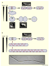
Roles for the actin (MreB) and tubulin (FtsZ) cytoskeletons in shaping E. coli cells. Depletion (vertical triangles) of only MreB (A) or only FtsZ (B) during growth disrupts each cytoskeleton and causes cells to either slowly become spheroidal (A) or form cylindrical filaments (B) over time (red downward arrows). Predivisional cells with normal levels of MreB (red) and FtsZ (green) have a Z ring surrounded by MreB rings (top rows in A and B). After cell growth and division over time (rightward black arrows), FtsZ and MreB relocalize into helical patterns, and prepare to divide at the next division site (yellow dashes). During MreB depletion (A), cells will form Z arcs (green) and divide, often asymmetrically, at those arcs to form a diplococcus, which then over time (rightward black arrows) separates to form a single round cell capable of dividing again in a perpendicular plane (yellow dashed line). These MreB-lacking cells will continue to grow and divide as spheroids. In contrast, FtsZ-depleted cells (B) continue to grow their sidewall and elongate their MreB cables but fail to divide, thus forming long filaments. The MreB double helix is shown as a single helix for simplicity. Micrographs representing some of the steps are shown next to the appropriate diagram.
Figure 3
Figure 3
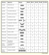
Phylogeny of some shape-determining proteins across representative bacteria. The species are grouped by family. For each species, typical cell shape is shown (not drawn to scale), along with the presence or absence of the protein as encoded in the genomic sequence. The presence or absence of MreB, MreC, MreD or RodZ in different species as identified by STRING COG was described in [61]. STRING COG was also used to identify DivIVA homologs. DivIVA is absent in all the Gram-negative species listed, but some members of the Gram-negative δ-proteobacteria contain DivIVA orthologs.
Figure 4
Figure 4
Cytoskeletons trigger new shapes in bacteria. Shown are two examples of new shapes resulting from bacterial cytoskeletal proteins. (A) In Streptomyces, DivIVA (orange balls) forms large assemblies at the poles of hyphae, possibly recognizing a region of sharp membrane curvature, but also forms seemingly random foci along the sidewall. As DivIVA tends to self-assemble, larger foci are easily generated (middle panel). These larger assemblages probably trigger branch formation, because a DivIVA focus usually forms under a new pole prior to visible branch initiation. A DivIVA lattice is subsequently maintained at the new branch tip (green) and is required for continued tip extension. (B) Cells of Mycoplasma pneumoniae lack peptidoglycan, and would be spheroidal without a cytoskeleton (top). Cytoskeletal proteins form the terminal organelle, which is important for gliding motility (red rightward arrow); the resulting cell extension (red) upon movement and subsequent duplication of the organelle at the opposite pole (not shown) gives these wall-less cells a roughly cylindrical shape with narrow tips.

Similar articles

Cited by

References

    1. Vollmer W, Blanot D, de Pedro MA. Peptidoglycan structure and architecture. FEMS Microbiol Rev. 2008;32:149–167. - PubMed
    1. den Blaauwen T, de Pedro MA, Nguyen-Disteche M, Ayala JA. Morphogenesis of rod-shaped sacculi. FEMS Microbiol Rev. 2008;32:321–344. - PubMed
    1. Cabeen MT, Jacobs-Wagner C. Skin and bones: the bacterial cytoskeleton, cell wall, and cell morphogenesis. J Cell Biol. 2007;179:381–387. - PMC - PubMed
    1. Park JT, Uehara T. How bacteria consume their own exoskeletons (turnover and recycling of cell wall peptidoglycan) Microbiol Mol Biol Rev. 2008;72:211–227. table of contents. - PMC - PubMed
    1. Young KD. The selective value of bacterial shape. Microbiol Mol Biol Rev. 2006;70:660–703. - PMC - PubMed

MeSH terms

LinkOut - more resources