. 2010 Apr;24(4):1043-55.
doi: 10.1096/fj.09-139634. Epub 2009 Nov 11.

RAGE-dependent signaling in microglia contributes to neuroinflammation, Abeta accumulation, and impaired learning/memory in a mouse model of Alzheimer's disease

Affiliations

RAGE-dependent signaling in microglia contributes to neuroinflammation, Abeta accumulation, and impaired learning/memory in a mouse model of Alzheimer's disease

Fang Fang et al. FASEB J. 2010 Apr.

Abstract

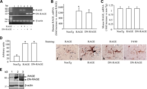
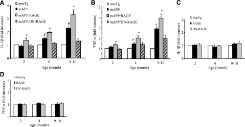
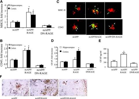
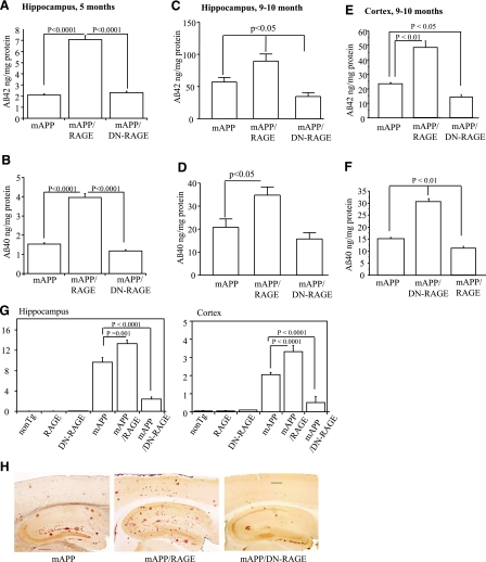
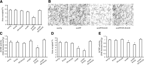
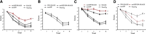
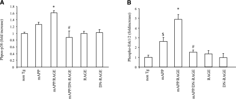
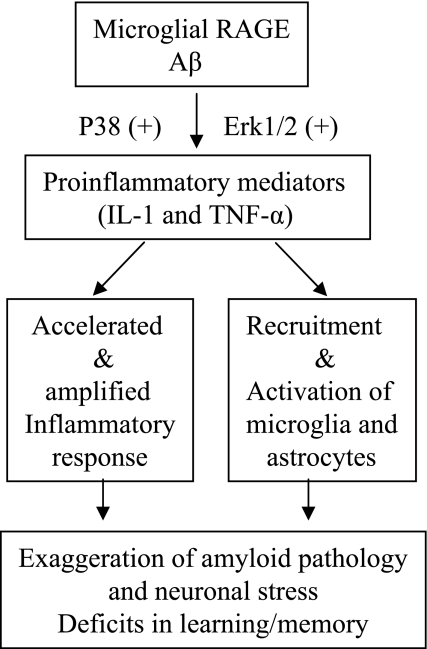
Microglia are critical for amyloid-beta peptide (Abeta)-mediated neuronal perturbation relevant to Alzheimer's disease (AD) pathogenesis. We demonstrate that overexpression of receptor for advanced glycation end products (RAGE) in imbroglio exaggerates neuroinflammation, as evidenced by increased proinflammatory mediator production, Abeta accumulation, impaired learning/memory, and neurotoxicity in an Abeta-rich environment. Transgenic (Tg) mice expressing human mutant APP (mAPP) in neurons and RAGE in microglia displayed enhanced IL-1beta and TNF-alpha production, increased infiltration of microglia and astrocytes, accumulation of Abeta, reduced acetylcholine esterase (AChE) activity, and accelerated deterioration of spatial learning/memory. Notably, introduction of a signal transduction-defective mutant RAGE (DN-RAGE) to microglia attenuates deterioration induced by Abeta. These findings indicate that RAGE signaling in microglia contributes to the pathogenesis of an inflammatory response that ultimately impairs neuronal function and directly affects amyloid accumulation. We conclude that blockade of microglial RAGE may have a beneficial effect on Abeta-mediated neuronal perturbation relevant to AD pathogenesis.-Fang, F., Lue, L.-F., Yan, S., Xu, H., Luddy, J. S., Chen, D., Walker, D. G., Stern, D. M., Yan, S., Schmidt, A. M., Chen, J. X., Yan, S. S. RAGE-dependent signaling in microglia contributes to neuroinflammation, Abeta accumulation, and impaired learning/memory in a mouse model of Alzheimer's disease.


Figures

Figure 1
Figure 1
Identification and characterization of RAGE and DN-RAGE mice. A) RT-PCR analysis of total RNA harvested from cerebral cortex of Tg RAGE mice (+, lanes 3–4), Tg DN-RAGE (+, lanes 5–6), and nonTg littermates (−, lanes 1–2) using primers for human RAGE. B, C) Quantitative real-time PCR for human (B) and mouse (C) RAGE mRNA in the brain of Tg mice. D, E) Immunoblotting of brain homogenates for RAGE. D) Quantification of RAGE immunoreactive bands in the brain of the indicated Tg mice. E) Top panel: representative immunoblots of brain homogenates from nonTg (lane 1), Tg RAGE (lane 2), and Tg DN-RAGE mice (lane 3). Bottom panel: immunoblots for β-actin used as protein-loading control. F–H) Immunostaining of brain sections from nonTg (F), Tg RAGE (G), and Tg DN-RAGE mice (H) for RAGE. I) Sequential staining of the section in panel H with F4/80 IgG (a microglial marker) shows an overlapping distribution of cells stained with antibodies to RAGE and F4/80. Sections were counterstained with hematoxylin. Above studies were performed on mice 3–4 mo of age.
Figure 2
Figure 2
Effect of microglial RAGE on induction of proinflammatory cytokines in brains of mAPP/RAGE and mAPP/DN-RAGE mice. Quantitative real-time PCR analysis of cytokines (IL-1β; A, C; and TNF-α; B, D) of total RNA extracted from cerebral cortex of the indicated Tg mice at 2, 4–5, and 9–10 mo of age (4–6 mice/group). There is no significant difference in IL-1β or TNF-α levels between nonTg and RAGE or DN-RAGE mice (P>0.01). #P < 0.01 vs. nonTg group; *P < 0.01 vs. other groups.
Figure 3
Figure 3
Effect of microglial RAGE on microgliosis and astrocytosis in brains of mAPP/RAGE and mAPP/DN-RAGE mice. A, B) Quantification of plaque-associated microglia positive for MHC II (A) or CD45 (B) in the cerebral cortex and hippocampus (5–6 mice/genotype). C) Representative sections from mAPP, mAPP/RAGE, and mAPP/DN-RAGE mice at 9–10 mo of age to demonstrate plaque-associated microglia. Arrows indicate cells reactive with microglial activation marker (MHC II or CD45; green). Amyloid plaques are red. D) Image analysis of plaque-associated astrocytosis in cortex and hippocampus of the indicated Tg mice at 9–10 mo of age (5–6 mice/genotype). E) Quantification of levels of GFAP in the cerebral cortex from the indicated Tg mice at 9–10 mo of age; n = 5–6 mice/group. *P < 0.05 vs. other groups; ELISA. F) Representative images from mAPP, mAPP/RAGE, and mAPP/DN-RAGE mice at 9–10 mo of age to demonstrate plaque-associated astrocytes. GFAP immunoreactive astrocyte clusters are black; amyloid plaques are brown.
Figure 4
Figure 4
Aβ levels and Aβ plaque load in brains of Tg mice. A–F) Aβ(1-40) and Aβ(1-42) levels were determined by ELISA in the hippocampus and cortex from mice of each of the genotypes at 5 mo (A, B) and 9–10 mo of age (C–F) (n=7–10 mice/group). G) Aβ deposits in hippocampus and neocortex were quantified by histological image analysis in the same group of mice after staining of brain sections with 3D6 antibody as indicated. H) Representative sections stained with 3D6 from Tg mAPP, Tg mAPP/MSR-RAGE, and Tg mAPP/MSR-DN-RAGE mice at 9–10 mo of age. Results are means ± se. Scale bar = 30 μm.
Figure 5
Figure 5
Effect of microglial RAGE on AChE activity of mAPP/RAGE and mAPP/DN-RAGE mice. A) AChE-positive neurites were visualized histochemically in the subiculum (Sb) of indicated genotypes at 4–5 mo of age. B) Representative images of AChE staining for indicated groups of mice at 4–5 mo of age. Scale bar = 5 μm. C) AChE activity in subiculum at 4–5 mo of age. D) AChE-positive neurites in subiculum at 9–10 mo of age. E) AChE activity in subiculum at 9–10 mo of age. n = 7–9 mice/group (A, B, D); 9–10 mice/group (C, E). *P < 0.01 vs. nonTg, RAGE, DN-RAGE, and mAPP/DN-RAGE groups; #P < 0.05 vs. nonTg, RAGE, DN-RAGE, and mAPP/RAGE groups. No significant differences were found among RAGE, DN-RAGE, nonTg, and mAPP/DN-RAGE groups.
Figure 6
Figure 6
Behavioral studies in mAPP/RAGE and mAPP/DN-RAGE mice. Spatial learning and memory were tested in the radial arm water maze at age 4–5 mo (A, B) and 9–10 mo (C, D) for the indicated mice; n = 7–9 male mice/genotype. Trials: 1–4, acquisition trials; R, retention trial. mAPP/RAGE mice demonstrated worsened spatial learning memory compared to nonTg mice and mAPP mice (A, C). Single mAPP mice at 9–10 mo of age showed impaired learning memory compared to nonTg littermate controls (C, D). mAPP/DN-RAGE mice significantly improved learning memory (D). *P < 0.01 (A, D), *P < 0.05 (C) vs. mAPP; #P < 0.01 vs. nonTg.
Figure 7
Figure 7
Effect of RAGE on activation of phosphorylation of p38 and ERK1/2 for in vivo mAPP mice. Phosphorylation of p38 (A) and ERK1/2 (B) in brain homogenates of indicated Tg mice was measured by ELISA; n = 3–8 mice/group. Vertical axis indicates fold-increased phosphorylated p38 (A) and phosphorylated ERK1/2 (B) (normalized to total p38 or ERK1/2, respectively) in the indicated Tg mice relative to nonTg mice. *P < 0.05 vs. nonTg, mAPP, RAGE, DN-RAGE, and mAPP/DN-RAGE groups; #P < 0.05 vs. mAPP group; $P < 0.05 vs. nonTg group.
Figure 8
Figure 8
Hypothesis for microglial RAGE involved in neuroinflammation, neuronal stress, and impaired learning/memory.

Similar articles

Cited by

References

    1. Uchihara T., Akiyama H., Kondo H., Ikeda K. Activated microglial cells are colocalized with perivascular deposits of amyloid-beta protein in Alzheimer’s disease brain. Stroke. 1997;28:1948–1950. - PubMed
    1. Giulian D., Li J., Leara B., Keenen C. Phagocytic microglia release cytokines and cytotoxins that regulate the survival of astrocytes and neurons in culture. Neurochem Int. 1994;25:227–233. - PubMed
    1. McGeer P. L., Rogers J., McGeer E. G. Inflammation, anti-inflammatory agents and Alzheimer disease: the last 12 years. J Alzheimers Dis. 2006;9:271–276. - PubMed
    1. Giulian D., Haverkamp L. J., Li J., Karshin W. L., Yu J., Tom D., Li X., Kirkpatrick J. B. Senile plaques stimulate microglia to release a neurotoxin found in Alzheimer brain. Neurochem Int. 1995;27:119–137. - PubMed
    1. Minghetti L. Role of inflammation in neurodegenerative diseases. Curr Opin Neurol. 2005;18:315–321. - PubMed

Publication types

MeSH terms

Substances